Internal Thoracic Artery to Vertebral Artery Bypass Surgery: A Cadaveric Feasibility Study.

World Neurosurg. 2019 Oct;130:e722-e725. doi: 10.1016/j.wneu.2019.06.203. Epub 2019 Jul 5.

Schmitt PJAltafulla JJKikuta SDupont GIwanaga JMonteith SLitvack ZDumont ASTubbs RS.


Abstract

OBJECTIVE:

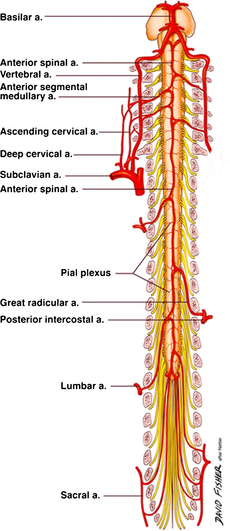
Posterior circulation strokes account for over one quarter of all ischemic strokes. The frequency of vertebral artery origin stenosis (VAOS) in patients with vertebrobasilar insufficiency (VBI) has been estimated to be as high 26%-32%, and VAOS is the direct cause of posterior circulation strokes in 9% of patients. This association could have a significant genetic component. This study examines the feasibility of the internal thoracic artery (ITA) as a donor vessel for revascularization in patients with VAOS.

METHODS:

Ten sides from 5 fresh-frozen white cadaveric necks derived from 3 women and 2 men were used in this study. The mean age of the cadavers at death was 77.2 years (range, 68-88 years). The subclavian artery, vertebral artery, and ITA were dissected. The length and diameter (proximal and distal) of the V1 segment and the length and diameter of the ITA were recorded. Finally, the ITA was transposed to the V1 segment of the vertebral artery (VA1).

RESULTS:

The mean length of the VA1 and its diameter at the proximal and distal parts were 35.51 and 3.69 mm, respectively. The mean length and diameter of the ITA were 26.53 and 3.27 mm, respectively. Rerouting the ITA to the VA1 was feasible without tension on all sides.

CONCLUSIONS:

This study indicates that the ITA is anatomically and hemodynamically an excellent option for bypass surgery in a VAOS scenario. We present convincing and reproducible data to aid neurosurgeons in choosing the procedure best suited to their patients.

Copyright © 2019 Elsevier Inc. All rights reserved.

KEYWORDS:

Internal mammary artery; Revascularization; Transposition; V1 segment; Vascular

Bilateral Lateral Costal Branches of the Internal Thoracic Arteries: A Case Report.

Kurume Med J. 2019 Sep 25;65(3):105-108. doi: 10.2739/kurumemedj.MS653003. Epub 2019 Aug 9.

Odo YIwanaga JTabira YWatanabe KSaga TTubbs RSFujishima YYamaki KI.


Abstract

We report a case of bilateral lateral costal branches (LCB) of the internal thoracic artery (ITA). On the left side, the ITA branched from the subclavian artery as a common trunk with the thyrocervical trunk. The left LCB flew into the collateral branch of the fifth intercostal artery after reaching the upper end of the sixth rib and after exiting the left ITA at the upper part of the first rib. The left ITA was disconnected near the second rib because it had been used for coronary artery bypass surgery. The right ITA arose from the anterior surface of the right subclavian artery just after the right ITA diverged from the brachiocephalic artery. The right LCB reached the upper end of the fifth rib and flew into the collateral branch of the fourth intercostal artery. The right ITA descended along the back of the costal cartilages as usual. The mechanism of the development of the LCB is thought to be due to a lateral longitudinal anastomosis connecting the inter-node arteries arising from the dorsal aorta during the embryonic phase. More anatomical and embryological studies are necessary to further elucidate this variant arterial branch.

KEYWORDS:

exertion angina; internal thoracic artery; intersegmental artery; lateral costal branch; longitudinal anastomosis

Pfeiffer type 2 syndrome: review with updates on its genetics and molecular biology.

Childs Nerv Syst. 2019 Sep;35(9):1451-1455. doi: 10.1007/s00381-019-04244-7. Epub 2019 Jun 21.

Rai RIwanaga JDupont GOskouian RJLoukas MOakes WJTubbs RS.


Abstract

INTRODUCTION:

Pfeiffer syndrome is a rare autosomal dominant inherited disorder associated with craniosynostosis, midfacial hypoplasia, and broad thumbs and toes. The syndrome has been divided into three clinical subtypes based on clinical findings.

METHODS:

This review will specifically examine the most severe type, Pfeiffer syndrome type 2, focusing on its genetics and molecular biology.

CONCLUSION:

This subtype of the syndrome is caused by de novo sporadic mutations, the majority of which occur in the fibroblast growth factor receptor type 1 and 2 (FGFR1/2) genes. There is not one specific mutation, however. This disorder is genetically heterogeneous and may have varying phenotypic expressions that in various cases have overlapped with other similar craniosynostoses. A specific missense mutation of FGFR2 causing both Pfeiffer and Crouzon syndromes has been identified, with findings suggesting that gene expression may be affected by polymorphism within the same gene. Compared to other craniosynostosis-related disorders, Pfeiffer syndrome is the most extreme phenotype, as the underlying mutations cause wider effects on the secondary and tertiary protein structures and exhibit harsher clinical findings.

KEYWORDS:

Acrocephalosyndactyly; Cloverleaf skull; Craniosynostosis; Fibroblast growth factor receptor (FGFR); Pfeiffer syndrome

Ventriculo-humeral shunt: a cadaveric feasibility study with application to treating hydrocephalus.

Childs Nerv Syst. 2019 Sep;35(9):1561-1564. doi: 10.1007/s00381-019-04175-3. Epub 2019 May 4.

Kassem MWIshak BMehta KIwanaga JLoukas MTubbs RS.


Abstract

PURPOSE:

Intraosseous vascular access is often used when vascular access is difficult. However, the use of this space for a receptacle for cerebrospinal fluid (CSF) diversion has been scantly considered.

MATERIALS AND METHODS:

Six upper limbs of fresh frozen cadavers were used for this study. In the supine position, a small hole was drilled through the outer cortex of the proximal humerus and into the medullary cavity. A 16-gauge needle was placed into the hole in the humerus and 150 cc of saline infused. Next, the adjacent axillary vein and tributaries were dissected to observe dilation or the presence of the blue-colored saline. For part two of the study, shunt tubing was passed subcutaneously from a supraclavicular incision to the hole made in the humerus. Range of motion of the shoulder was then performed.

RESULTS:

On all sides, all 150 cc of fluid was easily infused into the humerus. No specimen was found to have leakage from the drill hole site or into the extravascular soft tissues. With dissection of the axillary vein and its tributaries, all sides were found to have engorgement of these vessels. No tension was placed on the distal shunt tubing with full range of motion of the shoulder.

CONCLUSION:

Based on our study, the humerus is another option available to the neurosurgeon for CSF diversion.

KEYWORDS:

Humerus; Hydrocephalus; Intraosseous; Ventricular shunts

Marco antonio della torre and leonardo da vinci.

Clin Anat. 2019 Sep;32(6):744-748. doi: 10.1002/ca.23393. Epub 2019 May 17.

Picardi EEEMacchi VPorzionato ABoscolo-Berto RLoukas MTubbs RSDe Caro R.


Abstract

Thanks to collaborations with anatomists and the possibility of performing public dissections during the Renaissance period, artists started to represent the human body more accurately and objectively in their drawings. This cultural change provided the basis for subsequent advances in education and learning as well as the institution of an obligatory anatomy course in the Academies of Arts. The encounter in Pavia between the eclectic artist Leonardo Da Vinci (1452-1519) and the physician Marco Antonio Della Torre (1481-1511), Professor of Theoretical Medicine at the University of Padova, who near the end of his short life founded the "Anatomical School" of the University of Pavia, could be considered a perfect example of this collaboration. According to the historian Giorgio Vasari, while Della Torre practiced dissection, Leonardo made a book of drawings with red chalk annotated by pen. All these anatomical drawings by Leonardo, preserved at Windsor Castle as the property of the Queen of England, date from 1513, after the death of Della Torre. During the same period, Leonardo started his own dissections in the crypt of Santa Maria Nuova in Florence with his anatomical knowledge already mature and consolidated. The aim of the present study is to present the life of Della Torre, his intense scientific activity between Padova and Pavia, and his relationship with Leonardo Da Vinci. In particular, we found the only available manuscript of Della Torre in the Marciana library of Venice. Clin. Anat. 32:744-748, 2019.

© 2019 Wiley Periodicals, Inc.

KEYWORDS:

Leonardo Da Vinci; Marco Antonio Della Torre; anatomy; art; dissection

Neurological examination of the infant: A Comprehensive Review.

Clin Anat. 2019 Sep;32(6):770-777. doi: 10.1002/ca.23352. Epub 2019 Mar 8.

Salandy SRai RGutierrez SIshak BTubbs RS.


Abstract

Clinicians are required to perform neurological examinations on infants to ensure they meet developmental milestones. A full neurological examination includes evaluating motor and sensory function, assessing the status of the cranial nerves, testing primitive reflexes, and atypical responses to further evaluate any developmental pathologies. The difficulty in maintaining infants' cooperation requires resourcefulness on the clinician's part to understand what is being tested and in what way to complete the examination. This literature will provide clinicians with guidance on the way to conduct a thorough neurological examination of infants. Clin. Anat. 32:770-777, 2019.

© 2019 Wiley Periodicals, Inc.

KEYWORDS:

anatomy; cranial nerves; neurological examination; pediatric examination; physical exam; primitive; reflexes

The Basal Subarachnoid Cisterns: Surgical and Anatomical Considerations.

World Neurosurg. 2019 Sep;129:190-199. doi: 10.1016/j.wneu.2019.05.087. Epub 2019 May 25.

Altafulla JBordes SJenkins SLitvack ZIwanaga JLoukas MTubbs RS.


Abstract

The basal subarachnoid cisterns are expansions of the subarachnoid space and transmit cranial nerves and intracranial vessels. Providing neurosurgeons with key concepts, anatomical landmarks, and techniques can result in safer procedures and better patient outcomes. In this review, we discuss the major basal subarachnoid cisterns including their embryology, history, anatomical descriptions, and use during surgical approaches.

Copyright © 2019 Elsevier Inc. All rights reserved.

KEYWORDS:

Basal cisterns; Cerebrospinal fluid; Cranial nerves; Endoscopic; Third ventriculostomy

The ABC's of Spinal Decompression: Pearls and Technical Notes.

World Neurosurg. 2019 Sep;129:e146-e151. doi: 10.1016/j.wneu.2019.05.064. Epub 2019 May 16.

Ramey WLAltafulla JYilmaz EIshak BJack ALitvack ZNOskouian RJTubbs RSChapman JR.


Abstract

OBJECTIVE:

The foundation of spine surgery centers on the proper identification, decompression, and stabilization of bony and neural elements. We describe easily reproducible and reliable methods for optimal decompression and release of neural structures to alleviate symptoms and improve patients' quality of life.

METHODS:

Multiple spinal decompression techniques were described in procedures for which the goal of surgery was decompression alone or decompression and fusion. Eight fundamental techniques were described: inverted U-cut, J-cut, T-cut, L-cut, Z-cut, I-track cuts, C-cut, and O-cut.

RESULTS:

These foundational cuts may be combined, as needed, to develop an individually tailored approach to the patient's pathology.

CONCLUSIONS:

After properly identifying the anatomic structures, each of these techniques provides a consistent, reproducible, and efficient means to decompress the spine under various circumstances. These techniques provide surgical trainees with a framework for approaching surgical decompression.

Copyright © 2019 Elsevier Inc. All rights reserved.

KEYWORDS:

Bone cuts; Spinal decompression; Spine; Surgical technique

Intramuscular innervation of the lateral rectus muscle evaluated using sihler's staining technique: Potential application to strabismus surgery.

Clin Anat. 2019 Aug 20. doi: 10.1002/ca.23452. [Epub ahead of print]

Haładaj RWysiadecki GTubbs RS.


Abstract

The latest research suggests that the abducens nerve may be divided into subbranches that reach functionally distinct zones of the lateral rectus muscle. The goal of the study was to examine this muscle's innervation, including the detailed distribution of the intramuscular subbranches of the abducens nerve. Twenty-five lateral rectus muscle specimens were harvested (with the orbital segment of the abducens nerve), fixed in 10% formalin solution, and stained with Sihler's whole mount nerve staining technique. Subbranches running to the lateral rectus divided into two main groups: superior and inferior. Both groups of subbranches are distributed in a fan-shaped manner, show a characteristic "tree-like" branching pattern and form terminal plexus near the proximal half of the lateral rectus muscle. However, some smaller subbranches run as far as the muscle's insertion, and recurrent subbranches also reach its origin. With respect to their course to the muscle's origin or insertion, the smallest subbranches running within the muscle may be associated with innervation of the tendon. In the majority of cases (88%), superior and inferior subbranches of the abducens nerve overlapped in the central one-third of the lateral rectus muscle's width so that any clearly distinct anatomical segments of the muscle could be observed based on Sihler's technique. In the remaining 12% of specimens, superior and inferior groups of subbranches innervated two distinct compartments of the lateral rectus muscle with no overlapping. Dense, fan-shaped distribution of abducens nerve intramuscular subbranches can be observed within the lateral rectus muscle. Clin. Anat., 2019.

© 2019 Wiley Periodicals, Inc.

KEYWORDS:

Sihler's stain; abducens nerve; intramuscular nerve distribution; lateral rectus muscle; muscle compartmentalization

Craniometrics and Ventricular Access: A Review of Kocher's, Kaufman's, Paine's, Menovksy's, Tubbs', Keen's, Frazier's, Dandy's, and Sanchez's Points.

Oper Neurosurg (Hagerstown). 2019 Aug 16. pii: opz194. doi: 10.1093/ons/opz194. [Epub ahead of print]

Morone PJDewan MCZuckerman SLTubbs RSSinger RJ.


Abstract

Intraventricular access is frequently required during neurosurgery, and when neuronavigation is unavailable, the neurosurgeon must rely upon craniometrics to achieve successful ventricular cannulation. In this historical review, we summarize the most well-described ventricular access points: Kocher's, Kaufman's, Paine's, Menovksy's, Tubbs', Keen's, Frazier's, Dandy's, and Sanchez's. Additionally, we provide multiview, 3-dimensional illustrations that provide the reader with a novel understanding of the craniometrics associated with each point.

Copyright © 2019 by the Congress of Neurological Surgeons.

KEYWORDS:

Cerebrospinal fluid diversion; Dandy's point; External ventricular drain; Kocher's point; Paine's point; Ventricular access

Cervical spondylodiscitis caused by Candida albicans in a non-immunocompromised patient: A case report and review of literature.

Surg Neurol Int. 2019 Aug 2;10:151. doi: 10.25259/SNI_240_2019. eCollection 2019.

Huang SKappel ADPeterson CChamiraju PRajah GBMoisi MD.


Abstract

BACKGROUND:

Fungal cervical spondylodiscitis is rare and accounts for less than 1% of all cervical, thoracic, and lumbar vertebral osteomyelitis and discitis.

CASE DESCRIPTION:

A 32-year-old non-immunocompromised male presented with persistent neck pain and paresthesias. The magnetic resonance imaging of the cervical spine demonstrated a contrast-enhancing erosive lesion involving the cervical C6 and C7 vertebral bodies accompanied by epidural phlegmon. Blood culture was negative. The patient underwent a C6 and C7 anterior corpectomy with instrumented fusion (e.g., expandable cage C5 to T1). Intraoperatively, frank pus was noted within the C6-C7 disc space and was accompanied by thick prevertebral and epidural phlegmon extending from C5 to T1. Intraoperative cultures grew Candida albicans. Three days later, a C6-C7 laminectomy with C4-T2 posterior instrumented fusion was performed; the cultures again grew C. albicans. The patient was treated with intravenous micafungin for 14 days followed by 6-12 months of 400 mg oral fluconazole daily.

CONCLUSION:

There are few cases in literature where non-immunocompromised patients developed fungal cervical spondylodiscitis. Prompt diagnosis and appropriate management are critical to effectively treat these patients. Surgical intervention may warrant corpectomy, discectomy, and operative debridement followed by long-term targeted antifungal therapy.

KEYWORDS:

Candida albicans; Cervical spine; Discitis; Osteomyelitis; Spondylodiscitis

Anatomical Feasibility Study of the Digastric Branch of the Facial Nerve: A Potential Donor for Facial Nerve Reanimation.

J Oral Maxillofac Surg. 2019 Aug;77(8):1733.e1-1733.e6. doi: 10.1016/j.joms.2019.04.010. Epub 2019 Apr 18.

Kikuta S1, Iwanaga J2, Watanabe K3, Kusukawa J4, Tubbs RS5.


Abstract

PURPOSE:

The digastric branch is the second branch of the facial nerve after emerging from the stylomastoid foramen. To the best of our knowledge, few detailed anatomic studies of the digastric branch have been performed. Moreover, the use of the digastric branch for facial nerve reanimation has not been explored.

MATERIALS AND METHODS:

Nine sides of 5 fresh frozen cadavers were dissected for anatomic observation of the digastric branch and exploration of the feasibility of the digastric branch as a donor for facial nerve reanimation. The facial nerve trunk (FNT) and digastric branch were readily found by dissection using the tragal cartilage and tragal pointer as landmarks. The length and diameter of the digastric branch were measured, and the digastric branch was repositioned anterior to the FNT and the 2 major extracranial divisions of the FNT.

RESULTS:

On all sides, the digastric branch existed as a single branch and innervated only the posterior belly of the digastric muscle. The available length of the digastric branch was 13.28 ± 2.47 mm (range, 8.87 to 16.38 mm), and the mean diameter was 0.98 ± 0.31 mm (range, 0.64 to 1.64 mm). The digastric branch reached the FNT and its 2 major divisions without tension.

CONCLUSIONS:

The results of the present study have clarified the anatomic findings of the digastric branch in detail and the feasibility of using the digastric branch as a potential donor for facial nerve reanimation. The surgeon might consider the use of the digastric branch as a feasible donor for facial nerve reanimation based on the findings from our cadaveric study.

Copyright © 2019 American Association of Oral and Maxillofacial Surgeons. Published by Elsevier Inc. All rights reserved.

Guest UserComment
Artery of Adamkiewicz: a meta-analysis of anatomical characteristics.

Neuroradiology. 2019 Aug;61(8):869-880. doi: 10.1007/s00234-019-02207-y. Epub 2019 Apr 27..

Taterra DSkinningsrud BPękala PAHsieh WCCirocchi RWalocha JATubbs RSTomaszewski KAHenry BM.


Abstract

PURPOSE:

The artery of Adamkiewicz (AKA) provides the major blood supply to the anterior thoracolumbar spinal cord and iatrogenic injury or inadequate reconstruction of this vessel during vascular and endovascular surgery can result in postoperative neurological deficit due to spinal cord ischemia. The aim of this study was to provide comprehensive data on the prevalence and anatomical characteristics of the AKA.

METHODS:

An extensive search was conducted through the major electronic databases to identify eligible articles. Data extracted included study type, prevalence of the AKA, gender, number of AKA per patient, laterality, origin based on vertebral level, side of origin, morphometric data, and ethnicity subgroups.

RESULTS:

A total of 60 studies (n = 5437 subjects) were included in the meta-analysis. Our main findings revealed that the AKA was present in 84.6% of the population, and patients most frequently had a single AKA (87.4%) on the left side (76.6%) originating between T8 and L1 (89%).

CONCLUSION:

As an AKA is present in the majority of the population, caution should be taken during vascular and endovascular surgical procedures to avoid injury or ensure proper reconstruction. All surgeons operating in the thoracolumbar spinal cord should have a thorough understanding of the anatomical characteristics and surgical implications of an AKA.

KEYWORDS:

Adamkiewicz artery; Anatomy; Aortic aneurysm; Great anterior radiculomedullary artery; Thoracoabdominal aneurysm

Supernumerary incisive canals in a cadaver: a rare anatomical variation.

Surg Radiol Anat. 2019 Aug;41(8):977-978. doi: 10.1007/s00276-019-02191-1. Epub 2019 Jan 29.

Kikuta SIwanaga JKusukawa JTubbs RS.


Abstract

We present a rare anatomical variation in which an additional incisive canal was observed during the routine dissection of the palatal region in a cadaver. The diameter of the incisive and additional incisive canals measured 4.59 mm and 0.91 mm, respectively. Even though this variation is rare, oral surgeons should be aware of such anatomical variations for a better understanding.

KEYWORDS:

Anatomical variation; Anatomy; Incisive canal; Oral surgery; Palate

Early Complications and Cement Leakage in Elderly Patients Who Have Undergone (CT)-Guided Cement Augmented Pedicle Screw Placement.

World Neurosurg. 2019 Aug;128:e975-e981. doi: 10.1016/j.wneu.2019.05.047. Epub 2019 May 14.

Ishak BBajwa AASchneider TTubbs RSIwanaga JRamey WLUnterberg AWKiening KL.


Abstract

OBJECTIVE:

To assess early complications, mortality rate, and cement leakage in elderly patients who had undergone navigation-based pedicle screw placement of the thoracic and lumbar spine.

METHODS:

Eighty-six patients older than 65 years of age who had received cement-augmented pedicle screws for various conditions were retrospectively included between May 2008 and December 2016. Complications, mortality, and cement leakage were determined. All patients had a radiograph as a control. In patients with cement leakage seen on radiographs, a computed tomography scan of the surgical area was also obtained.

RESULTS:

Average age was 73.4 years (range 65-86 years). A total of 319 vertebral bodies with 637 screws were inserted, of which 458 screws were cement-augmented; 348 (76%) of the augmented screws were placed in the lumbar spine and 110 (24%) in the thoracic spine. Cement leakage occurred in 55 of 86 patients, of whom 52 (60%) were asymptomatic. In all cases with cement leakage (asymptomatic or symptomatic), cement could be found in the perivertebral veins: in the inferior vena cava in 25%, in the epidural space in 7%, in the azygos vein in 5%, and in pulmonary arteries in 7%.

CONCLUSIONS:

Our study confirms that the use of cement correlates with a high risk of cement leakage in elderly patients. Using computed tomography navigation for screw placement did not reduce the risk of venous cement leakage, but leakage into the epidural space or through a cortical defect seems to be low.

Copyright © 2019 Elsevier Inc. All rights reserved.

KEYWORDS:

Cement leakage; Complications; Elderly; Spinal instrumentation; Spinal navigation