Anatomical and Biomechanical Study of the Lumbar Interspinous Ligament.
Asian J Neurosurg. 2019 Nov 25;14(4):1203-1206. doi: 10.4103/ajns.AJNS_87_19. eCollection 2019 Oct-Dec.
Iwanaga J, Simonds E, Yilmaz E, Schumacher M, Patel M, Tubbs RS.
Abstract
OBJECTIVE:
The lumbar interspinous ligaments (ISLs) are thin and short fibers connecting adjacent spinous processes. However, their morphology is variably described and their biomechanics are not well understood. Therefore, the purpose of this study was to assess the anatomy and biomechanics of the lumbar ISL.
MATERIALS AND METHODS:
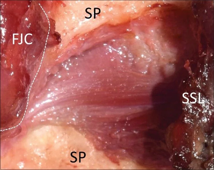
Five fresh frozen cadaveric specimens were dissected posteriorly to reveal and study the lumbar ISL. Measurements of the ligaments included the anterior vertical height (length A), the posterior vertical height (length P), and the length (length H) at each lumbar level. Next, 17 lumbar vertebral levels from 6 cadaveric specimens were used for tensile strength testing. The ISLs were subjected to vertically controlled increasing manual tension. The force necessary to disrupt the ISL was recorded.
RESULTS:
All the ISLs ran horizontally in an anterior-posterior direction with a slight curve. The average of length A, length P, and length H on the right sides was 9.82, 9.57, and 20.12 mm, respectively. The average of length A, length P, and length H on the left sides was 11.56, 12.01, and 21.42 mm, respectively. The mean tensile strength of the ISL was 162.33 (N) at L1/2, 85.67 (N) at L2/3, and 79 (N) at L3/4. There was a significant difference in the tensile force between L1/2 and L2/3 and L1/2 and L3/4 (P < 0.05). The ligaments became weaker with a descent along the lumbar levels.
CONCLUSION:
The results of this study might help surgeons understand pathology/trauma of the lumbar vertebral region.
Copyright: © 2019 Asian Journal of Neurosurgery.
KEYWORDS:
Biomechanics; cadaver; lumbar interspinous ligaments; spine; tensile strength