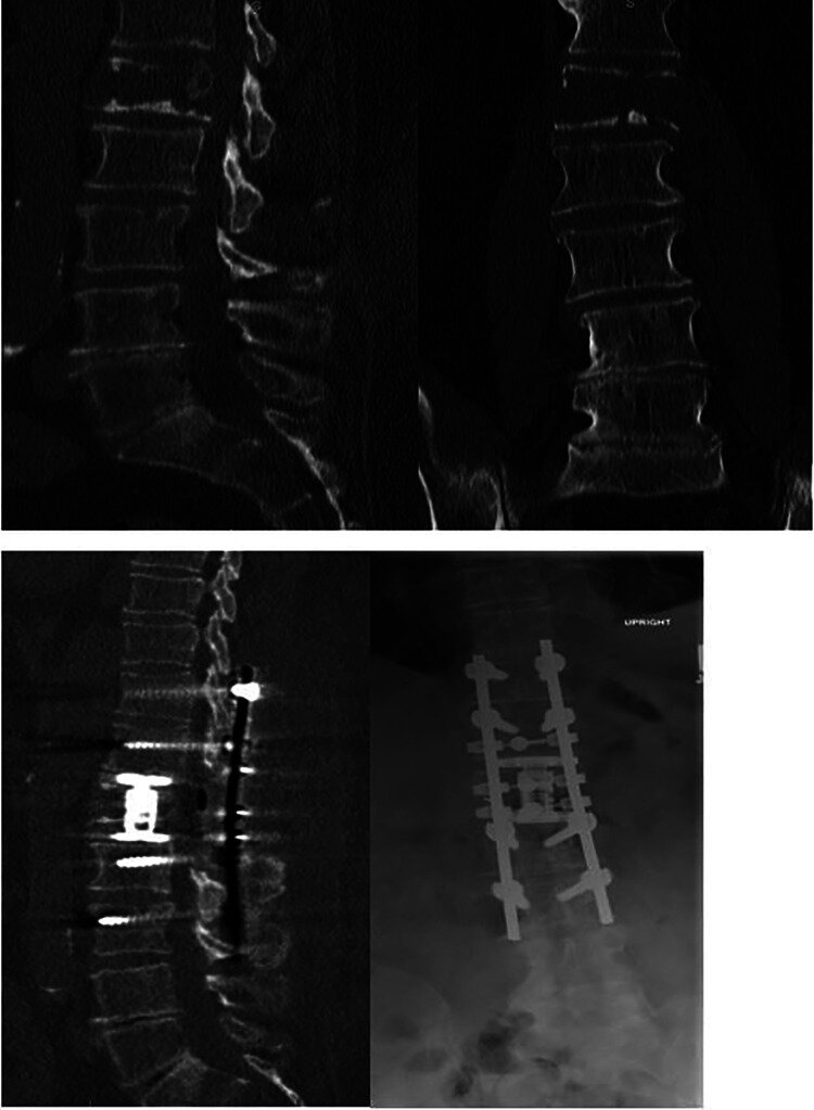
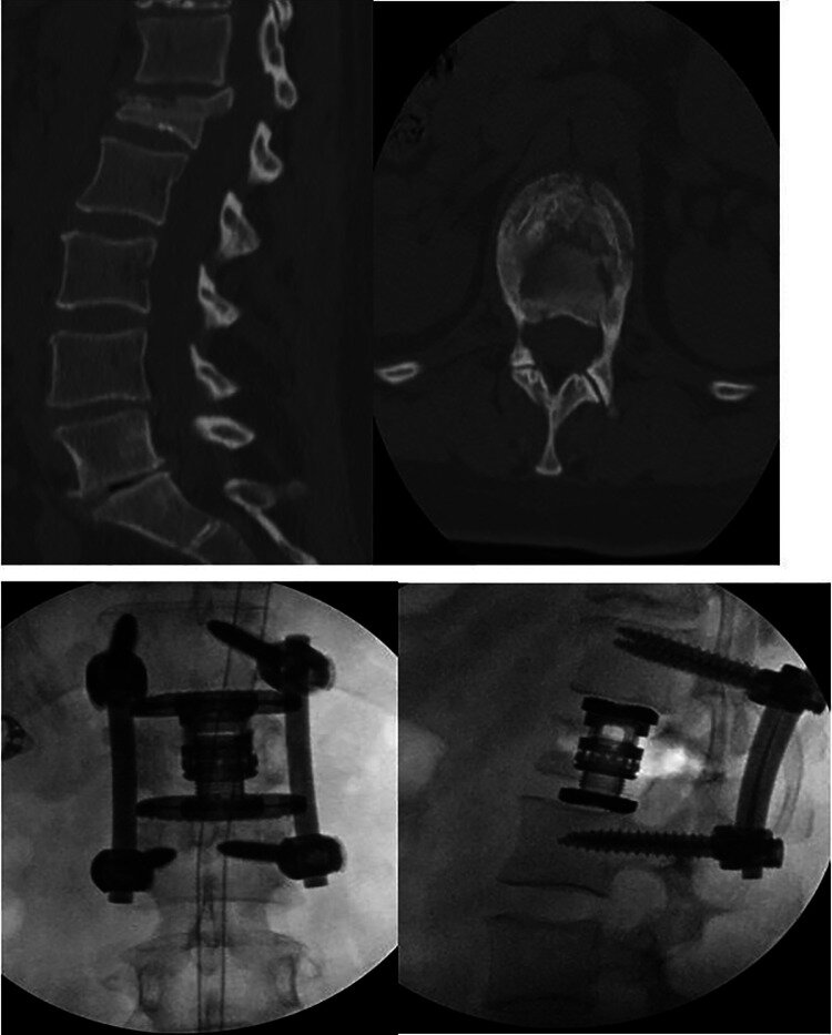
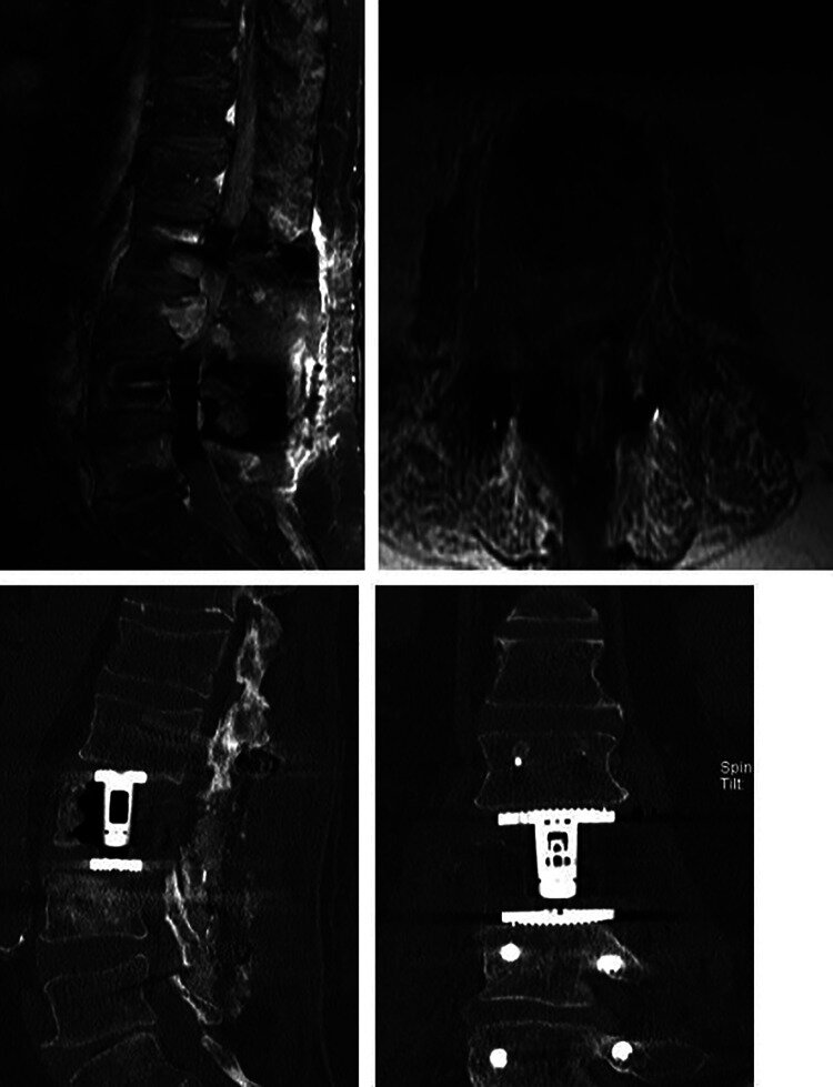
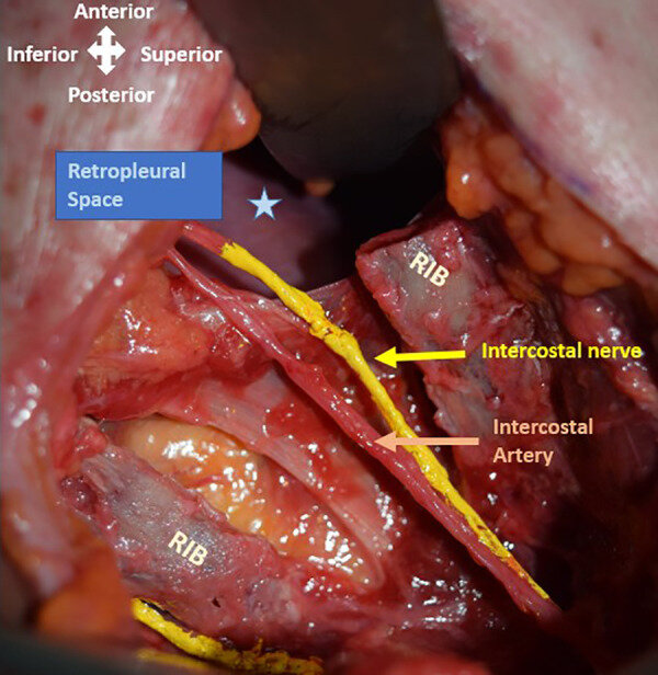
Extreme Lateral Interbody Fusion for Thoracic and Thoracolumbar Disease: The Diaphragm Dilemma

Global Spine J. 2021 May;11(4):515-524. doi: 10.1177/2192568220914883.Epub 2020 Mar 27.

Alexander Von Glinski, Christopher J Elia, Ariel Takayanagi, Emre Yilmaz, Basem Ishak, Joe Dettori, Benjamin A Schell, Erik Hayman, Clifford Pierre, Jens R Chapman, Rod J Oskouian


Abstract

Study design: Retrospective cohort study.

Objective: Complication profiles for lateral approaches to the spine are well established. However, the influence of level of surgery on complication rates and subtypes are less well established. To determine risk factors for complications as determined by level and surgery type in patients undergoing a lateral (retroperitoneal or retropleural approach) to the thoracolumbar spine.

Methods: All adult patients undergoing a lateral thoracolumbar fusion with or without posterior instrumentation performed at a single institution were identified. Primary outcomes assessed were presence of complication, complication subtype, and need for reoperation. The primary independent variables were spinal level (thoracic, thoracolumbar, or lumbar) and type of surgery (discectomy or corpectomy). Categorical outcomes were compared using chi-square test. Unadjusted and adjusted odds ratios for corpectomy status were calculated to determine risk of complication by level. P < .05 was considered statistically significant.

Results: A total of 165 patients aged 18 to 75 years were identified as having undergone a lateral fusion. Complication rates were 28.6%, 36.4%, and 11% for thoracic, thoracolumbar, and lumbar lateral approach fusions, respectively. Under univariate analysis, patients undergoing lateral approach in the thoracic spine group had significantly higher rates of postoperative complications than those in the lumbar group (P = .005). After adjusting for corpectomy status, there was no difference in complication rates.

Conclusions: Lateral (retroperitoneal or retropleural) approaches to the thoracic and thoracolumbar spine may be used with complication rates comparable to well-established lumbar approaches. Extent of surgery (corpectomy vs discectomy) rather than level of surgery may represent the primary driver of complications.

Keywords: complication; corpectomy; discectomy; lateral approach; neurologic deficit; pneumothorax.

Tyler LawsComment
Who Needs Surgical Stabilization for Pyogenic Spondylodiscitis? Retrospective Analysis of Non-Surgically Treated Patients

Global Spine J. 2021 Sep 16;21925682211039498. doi: 10.1177/21925682211039498. Online ahead of print.

Ronen Blecher, Sven Frieler, Bilal Qutteineh, Clifford A Pierre, Emre Yilmaz, Basem Ishak, Alexander Von Glinski, Rod J Oskouian, Moti Kramer, Michael Drexler, Jens R Chapman


Abstract

Study design: Retrospective case series analysis.

Objective: To identify relevant clinical and radiographic markers for patients presenting with infectious spondylo-discitis associated with spinal instability directly related to the infectious process.

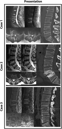
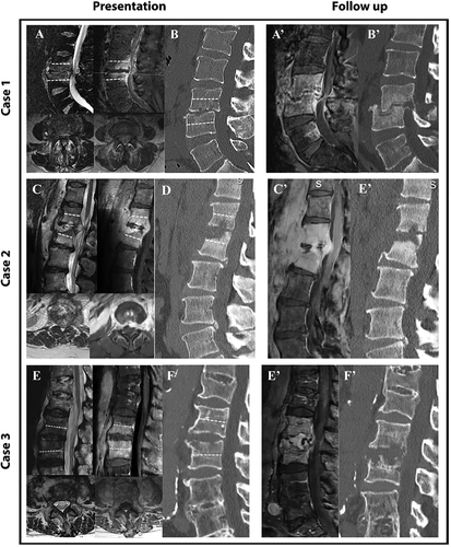
Methods: We evaluated patients presenting with de-novo intervertebral discitis or vertebral osteomyelitis /discitis (VOD) who initiated non-surgical treatment. Patients who failed conservative treatment and required stabilization surgery within 90 days were defined as "failed treatment group" (FTG). Patients who experienced an uneventful course served as controls and were labeled as "nonsurgical group" (NSG). A wide array of baseline clinical and radiographic parameters was retrieved and compared between 2 groups.

Results: Overall 35 patients had initiated non-surgical treatment for VOD. 25 patients had an uneventful course (NSG), while 10 patients failed conservative treatment ("FTG") within 90 days. Factors found to be associated with poorer outcome were intra-venous drug abuse (IVDA) as well as the presence of fever upon initial presentation. Radiographically, involvement of the same-level facets and the extent of caudal and rostral VB involvement in both MRI and CT were found to be significantly associated with poorer clinical and radiographic outcome.

Conclusions: We show that clinical factors such as IVDA status and fever as well as the extent of osseous and posterior element involvement may prove to be helpful in favoring surgical treatment early on in the management of spinal infections.

Keywords: infection; spondylitis; spondylodiscitis.


Tyler LawsComment
Surgical Management of Charcot Spinal Arthropathy in the Face of Possible Infection

Abstract

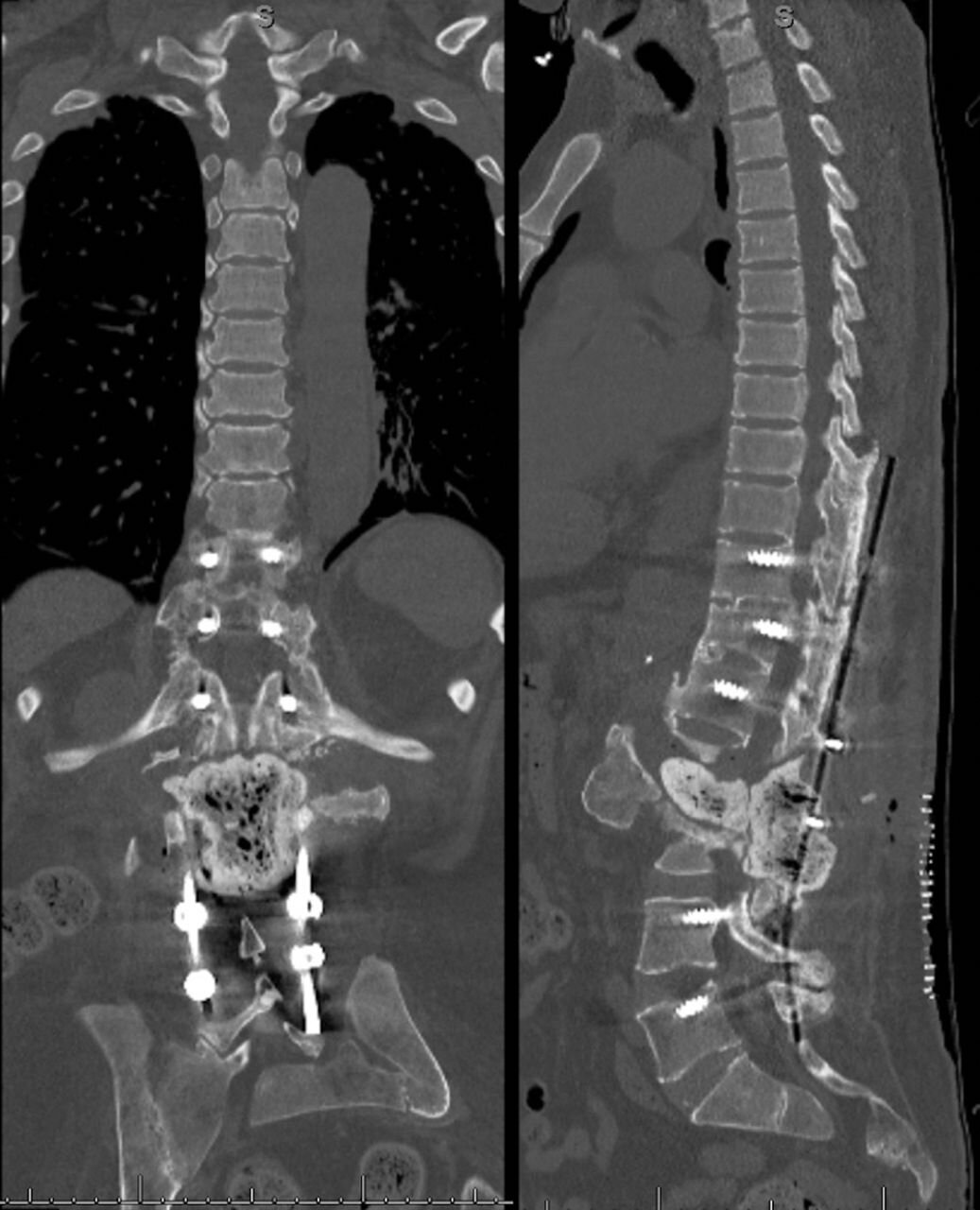
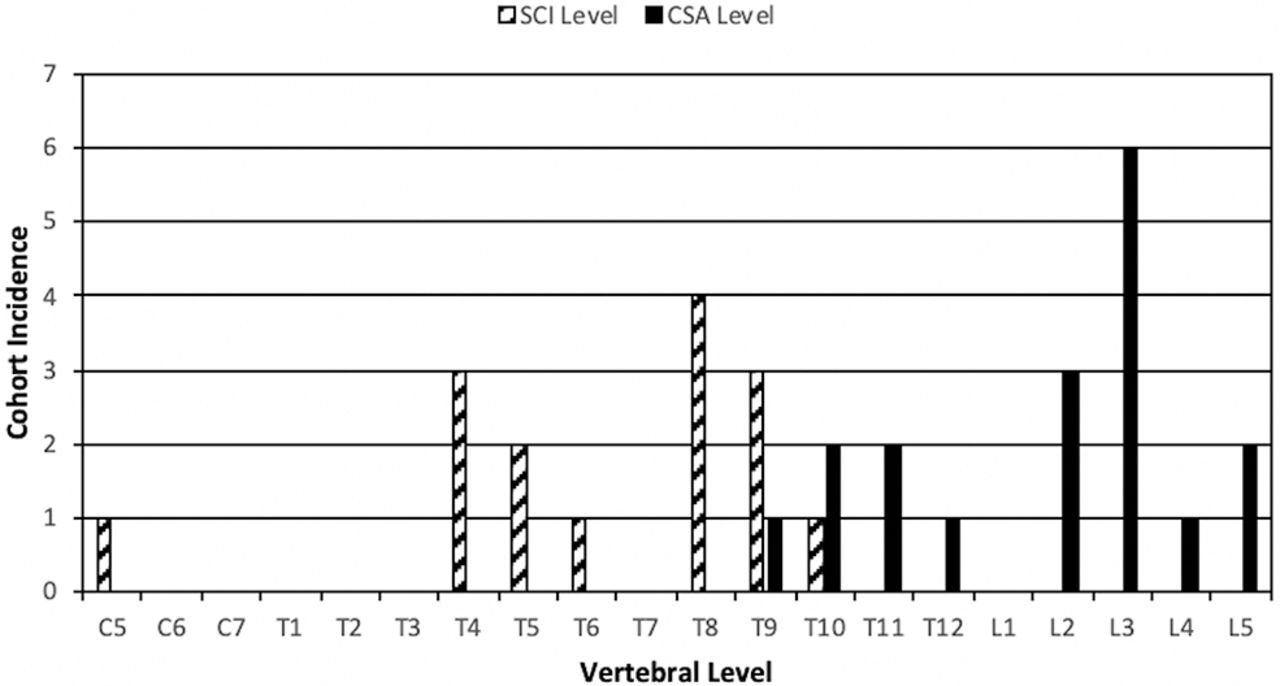
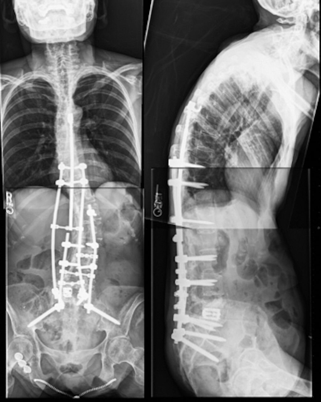
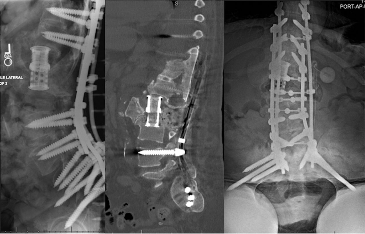
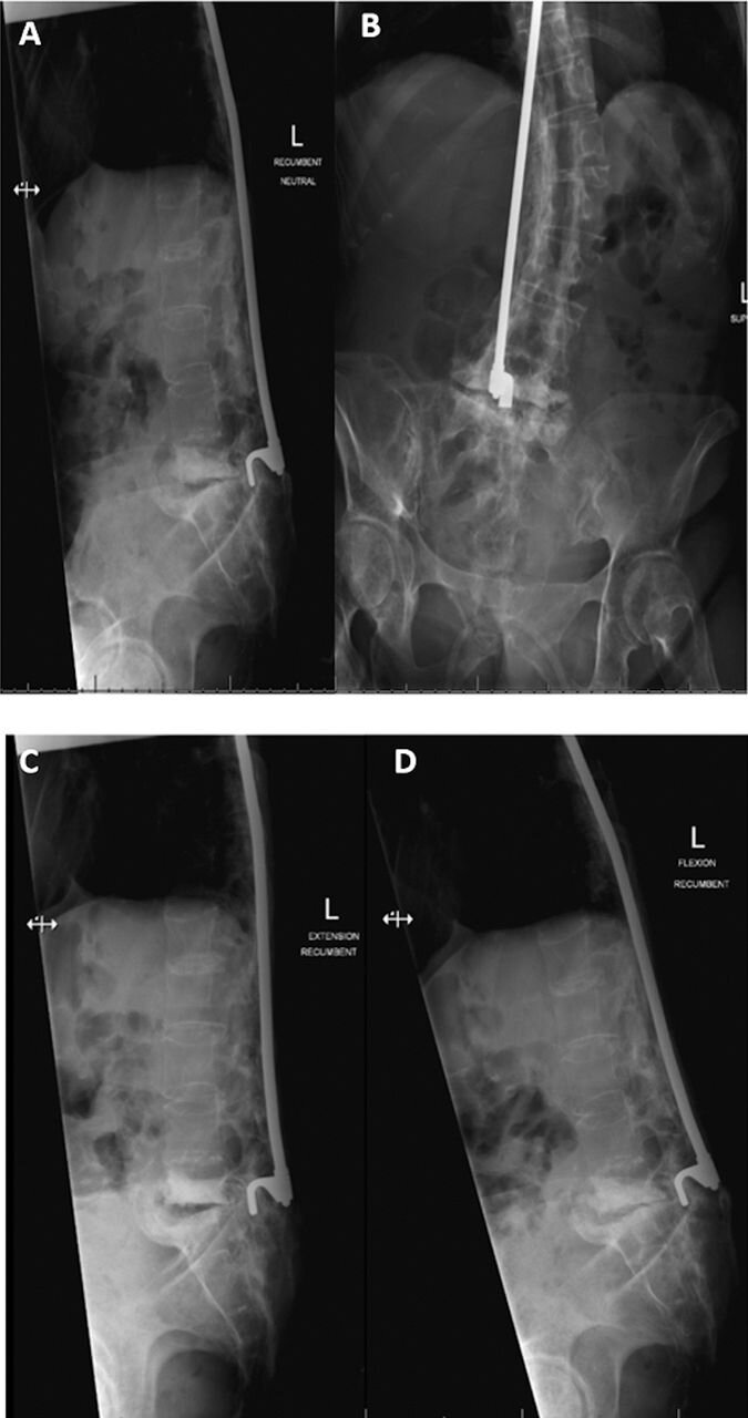
Background: The design is a retrospective cohort study. Charcot spinal arthropathy (CSA) is a rare and poorly understood progressive destructive spine condition that usually affects patients with preexisting spinal cord injury. The complexity of this condition, especially when additionally burdened by superimposed infection in the CSA zone, can potentially lead to suboptimal management such as protracted antibiotic therapy, predisposition to hardware failure, and pseudarthrosis. While in noninfected CSA primary stabilization is the major goal, staged surgical management has not been stratified based upon presence of a superinfected CSA. We compare clinical and radiological outcomes of surgical treatment in CSA patients with and without concurrent spinal infections.

Methods: Our single-institution database was reviewed for all patients diagnosed with CSA and surgically treated, who were subsequently divided into 2 cohorts: spinal arthropathy with superimposed infection and those without. Those were comparatively studied for complications and reoperation rate.

Results: Fifteen patients with CSA underwent surgical intervention; mean follow up of 15.3 months (range, 0-43). Eleven patients received stabilization with a quadruple-rod thoracolumbopelvic construct, while 4 patients with superinfected CSA underwent a staged procedure. Patients treated with a staged approach experienced fewer intraoperative complications (0% versus 18%) and fewer revision surgeries (25% versus 36%). Both cohorts had the same eventual healing.

Conclusions: Surgical management in CSA patients with primary emphasis on stability and modified surgical treatment based on presence of an active infection in the zone of neuropathic destruction will lead to similar eventual successful results with relatively few and manageable complications in this challenging patient population.

Level of evidence: 4.

Clinical relevance: The proposed treatment algorithm including the use of a quadruple-rod construct with lumbopelivic fixation and a staged approach in patients with superinfected CSA represents a reasonable option in the surgical treatment of CSA.

Keywords: Hounsfield unit (HU); bone morphogenic protein (BMP); polymethylmethacrylate (PMMA); quadruple-rod construct.


Tyler LawsComment
Three-stage revision arthroplasty for the treatment of fungal periprosthetic joint infection: outcome analysis of a novel treatment algorithm : a prospective study

Bone Jt Open. 2021 Aug;2(8):671-678. doi: 10.1302/2633-1462.28.BJO-2021-0002.R2.

Hinnerk Baecker, Sven Frieler, Jan Geßmann, Stephan Pauly, Thomas A Schildhauer, Yannik Hanusrichter


Abstract

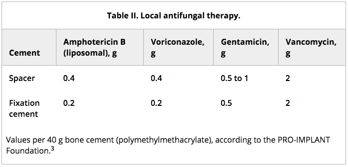
Aims: Fungal periprosthetic joint infections (fPJIs) are rare complications, constituting only 1% of all PJIs. Neither a uniform definition for fPJI has been established, nor a standardized treatment regimen. Compared to bacterial PJI, there is little evidence for fPJI in the literature with divergent results. Hence, we implemented a novel treatment algorithm based on three-stage revision arthroplasty, with local and systemic antifungal therapy to optimize treatment for fPJI.

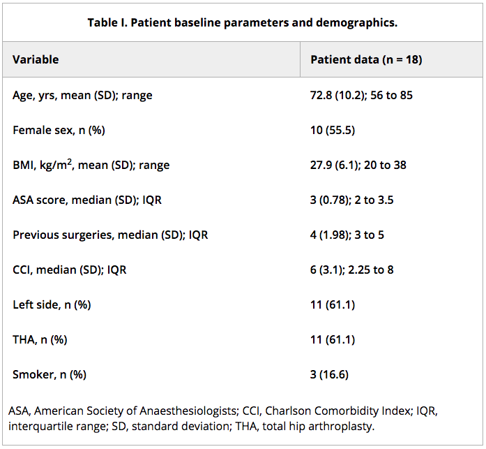
Methods: From 2015 to 2018, a total of 18 patients with fPJI were included in a prospective, single-centre study (DKRS-ID 00020409). The diagnosis of PJI is based on the European Bone and Joint Infection Society definition of periprosthetic joint infections. The baseline parameters (age, sex, and BMI) and additional data (previous surgeries, pathogen spectrum, and Charlson Comorbidity Index) were recorded. A therapy protocol with three-stage revision, including a scheduled spacer exchange, was implemented. Systemic antifungal medication was administered throughout the entire treatment period and continued for six months after reimplantation. A minimum follow-up of 24 months was defined.

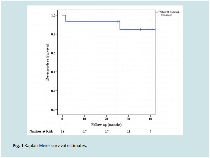
Results: Eradication of infection was achieved in 16 out of 18 patients (88.8%), with a mean follow-up of 35 months (25 to 54). Mixed bacterial and fungal infections were present in seven cases (39%). The interval period, defined as the period of time from explantation to reimplantation, was 119 days (55 to 202). In five patients, a salvage procedure was performed (three cementless modular knee arthrodesis, and two Girdlestone procedures).

Conclusion: Therapy for fPJI is complex, with low cure rates according to the literature. No uniform treatment recommendations presently exist for fPJI. Three-stage revision arthroplasty with prolonged systemic antifungal therapy showed promising results. Cite this article: Bone Jt Open 2021;2(8):671-678.

Keywords: BMI; Charlson Comorbidity Index (CCI); Fungal infection; Fungal prosthetic joint infection; Girdlestone procedures; Infection treatment; Joint Infection; PJI; Prosthetic joint infection; Revision arthroplasty; Total hip arthroplasty; Total knee arthroplasty; bone cement; fPJI; fungal infections; infections; knee arthrodesis; periprosthetic joint infections (PJI); revision joint arthroplasty.

Tyler Laws Comment
Iliac Screw Fixation Revisited: Improved Clinical and Radiologic Outcomes Using a Modified Iliac Screw Fixation Technique

Abstract

Study design: A retrospective study.

Objective: To describe the modified iliac screw (mILS) technique and compare it to other spinopelvic fixation techniques in terms of wound healing complications, hardware prominence, and failure.

Summary of background data: The traditional entry point of an iliac screw often causes postoperative gluteal pain from the prominent screw head. The use of an offset connector also adds a point of weakness to the construct. By choosing a different screw entry point offset connectors can be avoided, and the screw head itself is less prominent, thereby reducing postoperative discomfort.

Materials and methods: A retrospective analysis was performed of adult patients undergoing lumbopelvic fixation (LPF) between January 2014 and June 2019. Patients were grouped into 1 of 3 groups based on the technique of pelvic fixation: S2 alar-iliac (S2AI) screw, traditional iliac screw (tILS), and mILS. The primary outcome parameter was the minimal distance from screw head to skin. Secondary outcome parameters were instrumentation loosening/failure, adjacent level fractures, pseudoarthrosis, and medial or lateral iliac screw perforation.

Results: A total of 190 patients undergoing LPF were included in the following 3 groups: mILS group (n=113), tILS group (n=40), and S2AI group (n=37). The mean minimal distance from screw head to skin in the mILS group was 31.3 mm compared with 23.7 mm in the tILS group (P<0.00199). No statistically significant differences were found when comparing the 3 groups with respect to complications. The mILS group did not show any cases of prominent instrumentation and had the lowest rate of instrumentation failure.

Conclusions: The mILS technique is an acceptable alternative for LPF, offering the benefits of iliac screw fixation while avoiding offset connectors and screw prominence complications associated with tILS.

Level of evidence: Level III.


Tyler LawsComment
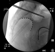
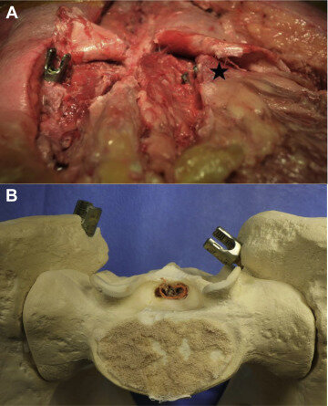
The Modified Iliac Screw: An Anatomic Comparison and Technical Guide

World Neurosurg. 2020 Apr;136:e608-e613. doi: 10.1016/j.wneu.2020.01.091.Epub 2020 Jan 23.

Alexander von Glinski, Emre Yilmaz, Basem Ishak, Erik Hayman, Wyatt Ramey, Andrew Jack, Joe Iwanaga, Rod J Oskouian, R Shane Tubbs, Jens R Chapman


Abstract

Background: Iliac screws are a widely used sacropelvic fixation technique, which is often criticized for its impaired wound healing owing to hardware prominence. The aim of this study was to present a modified iliac screw (MIS) fixation technique that uses a different entry point more medially and caudally to the posterior superior iliac spine next to the rudimentary S1-S2 joint. Soft tissue coverage and midline distance in an MIS and a traditional iliac screw were compared.

Methods: Two different variations of iliac screws were placed into 12 fresh frozen adult cadavers (9 male, 3 female, mean age at death 77.08 years, mean body mass index 23.4). The distance between the midline and the center of the screw head was measured. We also compared the angulation of the trajectories. After wound closure, we measured the distance between the iliac screw head and the skin.

Results: The mean distance from the screw tulip head to the skin was 2.43 cm (range, 1.2-4.2 cm) with the traditional iliac screw and 3.16 cm (range, 1.7-4.3 cm) with the MIS. The mean distance to the midline with the MIS was 3.1 cm (range, 2.4-4.5 cm) lateral to the midline compared with the traditional iliac screw, of which the mean was 4.2 cm lateral to the midline (range, 3.7-4.9 cm). Mean angulation was 10°.

Conclusions: The MIS avoids the use of connectors and provides less prominent pelvic fixation. Clinically, this might help prevent prominent hardware and related wound healing impairment.

Keywords: Iliac screws; Lumbar pelvic fixation; Modified iliac screw; Screw prominence; Soft tissue coverage.

Copyright © 2020 Elsevier Inc. All rights reserved.


Tyler LawsComment
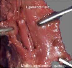
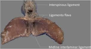
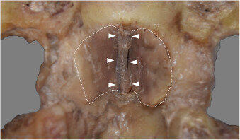
Discovery of a new ligament of the lumbar spine: the midline interlaminar ligament

Spine J. 2020 Jul;20(7):1134-1137. doi: 10.1016/j.spinee.2019.12.003.Epub 2019 Dec 9.

Emily Simonds, Joe Iwanaga, Basem Ishak, Miguel Angel Reina, Rod J Oskouian, R Shane Tubbs


Abstract

Background: The ligaments of the lumbar spine are integral to structural integrity and have been well-studied. However, during the routine dissection of the lumbar spine, we identified to our knowledge, a previously undescribed midline ligament near the ligamenta flava but distinctly separate.

Purpose: The purpose of this study was to investigate the morphology of this ligament termed by us the midline interlaminar ligament.

Study design: Cadaveric study.

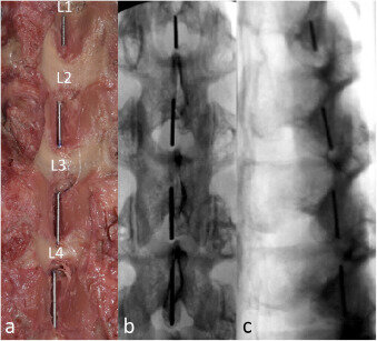
Methods: Thirty-four lumbar vertebral levels from 10 fresh frozen adult cadavers were dissected. The ligamenta flava were dissected anteriorly. The junction between the right and left ligamenta flava was observed and the presence and morphometrics of the median interlaminar ligament (MIL) were recorded. Random ligaments underwent tensile strength testing. Metal wires were also placed on the ligaments and fluoroscopy performed.

Results: Twenty-six out of 34 (76.5%) lumbar levels were found to have a MIL traveling on the internal aspect of the most medial aspect of the laminae and positioned slightly anterior to the plane of the ligamenta flava. The mean length and width of the MIL were 9.03±4.29 mm and 4.94±1.56 mm, respectively. The mean force necessary until failure for the MIL was 12.3N.

Conclusions: Based on our findings, a distinct MIL was identified in the lumbar spine at the majority of lumbar levels.

Clinical significance: The MIL might have clinical significance and potential biomechanical importance. Further studies are now necessary to better elucidate this anatomical structure.

Keywords: Anatomy; Cadaver; Ligamenta flava; Lumbar spine; Spine ligaments; Spine surgery.

Copyright © 2019 Elsevier Inc. All rights reserved.


Tyler LawsComment
Factors Associated With C5 Palsy Following Cervical Spine Surgery: A Systematic Review

Global Spine J. 2019 Dec;9(8):881-894. doi: 10.1177/2192568219874771.Epub 2019 Nov 22.

Andrew Jack, Wyatt L Ramey, Joseph R Dettori, Zane A Tymchak, Rod J Oskouian, Robert A Hart, Jens R Chapman, Dan Riew


Abstract

Study design: Systematic review.

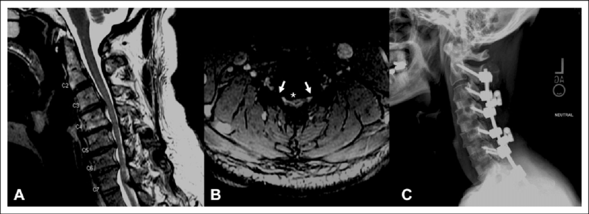
Objectives: C5 palsy (C5P) is a not uncommon and disabling postoperative complication with a reported incidence varying between 0% and 30%. Among others, one explanation for its occurrence includes foraminal nerve root tethering. Although different risk factors have been reported, controversy about its causation and prevention persists. Inconsistent study findings contribute to the persistent ambiguity leading to an assumption of a multifactorial nature of the underlying C5P pathophysiology. Here, we report the results of a systematic review on C5P with narrow inclusion criteria in the hope of elucidating risk factors for C5P due to a common pathophysiological mechanism.

Methods: Electronic databases from inception to March 9, 2019 and references of articles were searched. Narrow inclusion criteria were applied to identify studies investigating demographic, clinical, surgical, and radiographic factors associated with postoperative C5P.

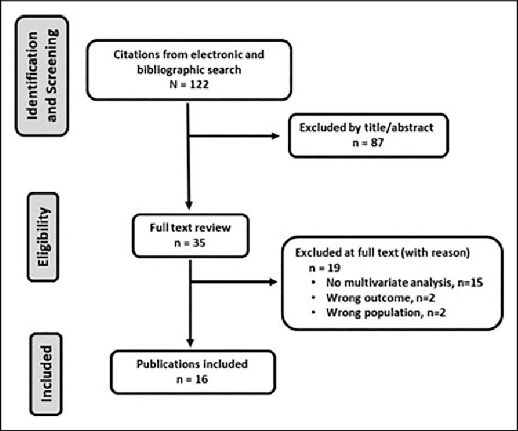
Results: Sixteen studies were included after initial screening of 122 studies. Eighty-four risk factors were analyzed; 27 in ≥2 studies and 57 in single studies. The pooled prevalence of C5P was 6.0% (range: 4.2%-24.1%) with no consistent evidence that C5P was associated with demographic, clinical, or specific surgical factors. Of the radiographic factors assessed, specifically decreased foraminal diameter and preoperative cord rotation were identified as risk factors for C5P.

Conclusion: Although risk factors for C5P have been reported, ambiguity remains due to potentially multifactorial pathophysiology and study heterogeneity. We found foraminal diameter and cord rotation to be associated with postoperative C5P occurrence in our meta-analysis. These findings support the notion that factors contributing to, and acting synergistically with foraminal stenosis increase the risk of postoperative C5P.

Keywords: C5 palsy; cervical spine surgery; complications; myelopathy.

© The Author(s) 2019.


Tyler LawsComment
Intraoperative Ischemic Stroke in Elective Spine Surgery: A Retrospective Study of Incidence and Risk

Spine (Phila Pa 1976). 2020 Jan 15;45(2):109-115. doi: 10.1097/BRS.0000000000003184.

Basem Ishak, Amir Abdul-Jabbar, Amit Singla, Emre Yilmaz, Alexander von Glinski, Wyatt L Ramey, Ronen Blecher, Zane Tymchak, Rod Oskouian, Jens R Chapman


Abstract

Study design: . Retrospective study.

Objective: . To determine incidence, risk factors, complications, and early postoperative outcome in patients with intraoperative ischemic stroke during elective spine surgery.

Summary of background data: . Overall, stroke is the fifth leading cause of death in the United States and the second leading cause of death worldwide. It can be a catastrophic event and the main cause of neurological disability in adults.

Methods: . A retrospective review of the electronic medical records of patients who underwent elective spine surgery between January 2016 and November 2018 at a larger tertiary referral center was conducted. Patients with infection and neoplastic disease were excluded. Patient demographics, pre- and postoperative neurological status, surgical treatment, surgical time, blood loss, intraoperative abnormalities, risk factors, history of stroke, medical treatment, diagnostics, hospital stay, complications, and mortality were collected.

Results: . Out of 5029 surgically treated patients receiving elective spine surgery, a total of seven patients (0.15%) were identified who developed an ischemic stroke during the surgical procedure. Patients were predominantly females (n = 6). Ischemic pontine stroke occurred in two patients. Further distributions of ischemic stroke were: left caudate nucleus, left posterior inferior cerebellar artery, left external capsule, left middle cerebral artery, and acute ischemic supratentorial spots. The main risk factors identified for intraoperative ischemic stroke include hypertension, diabetes, smoking, dyslipidemia, and possibly major intraoperative CSF leak. Three patients (43%) had neurological deficits which did not improve during hospital stay. Two patients recovered fully and two patients died. Therefore, in-hospital mortality rate of this subset of patients was 29%.

Conclusion: . With the increase of spinal procedures, it is important to identify patients at risk for having an ischemic stroke and to optimize their comorbidities preoperatively. Patients with intraoperative ischemic stroke carry a higher risk for morbidity and mortality during the index hospitalization.

Level of evidence: 4.


Tyler LawsComment
Risk Factors Associated with 90-day Readmissions Following Odontoid Fractures- A Nationwide Readmissions Database Study

Spine (Phila Pa 1976). 2021 Feb 22. doi: 10.1097/BRS.0000000000004010.Online ahead of print.

Alexander von Glinski, Sven Frieler, Christopher Elia, Tye Patchana, Ariel Takayanagi, Varun Arvind, Clifford Pierre, Basem Ishak, Jens Chapman, Rod Oskouian


Abstract

Study design: Nationwide Readmissions Database Study.

Objective: To investigate readmission rates and factors related to readmission after surgical and non-surgical management of odontoid fractures.

Summary of background data: Management of odontoid fractures, which are the most common isolated spine fracture in the elderly, continues to be debated. The choice between surgical or non-surgical treatment has been reported to impact mortality and might influence readmission rates. Hospital readmissions represent a large financial burden upon our healthcare system. Factors surrounding hospital readmissions, would benefit from a better understanding of their associated causes in order to lower health care costs.

Methods: A retrospective study was performed using the 2016 Healthcare Utilization Project (HCUP) Nationwide Readmission Database (NRD). Demographic information and factors associated with readmission were collected. Readmission rates, complications, length of hospital stay were collected. Patients treated operatively, non-operatively, and patients who were readmitted or not readmitted were compared. Statistical analysis was performed using open source software SciPy (Python v1.3.0) for all analyses.

Results: We identified 2,921 patients who presented with Type II dens fractures from January 1st 2016 to September 30th 2016, 555 of which underwent surgical intervention. The readmission rate in patients who underwent surgery was 16.4% (91/555) and 29.4% (696/2366) in the non-operative group. Hospital costs for readmitted and non-readmitted patients were $353,704 and $174,922, and $197,099 and $80,715 for non-operatively managed patients, respectively. Medicaid and Medicare patients had the highest readmission rate in both groups. Charlson and Elixhauser comorbidity indices were significantly higher in patients who were readmitted (p < 0.0001).

Conclusion: We report an overall 90-day readmission rate of 16.4% and 29.4%, in operative and non-operative management of type II odontoid fractures, respectively. In the face of a rising incidence of this fracture in the elderly population, an understanding of the comorbidities and age-related demographics associated with 90-day readmissions following both surgical and non-surgical treatment are critical.Level of Evidence: 3.


Tyler LawsComment
Contact Sports as a Risk Factor for Amyotrophic Lateral Sclerosis: A Systematic Review

Global Spine J. 2019 Feb;9(1):104-118. doi: 10.1177/2192568218813916.Epub 2019 Jan 31.

Ronen Blecher, Michael A Elliott, Emre Yilmaz, Joseph R Dettor, Rod J Oskouian, Akil Patel, Andrew Clarke, Mike Hutton, Robert McGuire, Robert Dunn, John DeVine, Bruce Twaddle, Jens R Chapman


Abstract

Study design: Systematic review.

Introduction: Amyotrophic lateral sclerosis (ALS) is a progressive neurodegenerative disease, ultimately resulting in paralysis and death. The condition is considered to be caused by a complex interaction between environmental and genetic factors. Although vast genetic research has deciphered many of the molecular factors in ALS pathogenesis, the environmental factors have remained largely unknown. Recent evidence suggests that participation in certain types of sporting activities are associated with increased risk for ALS.

Objective: To test the hypothesis that competitive sports at the highest level that involve repetitive concussive head and cervical spinal trauma result in an increased risk of ALS compared with the general population or nonsport controls.

Methods: Electronic databases from inception to November 22, 2017 and reference lists of key articles were searched to identify studies meeting inclusion criteria.

Results: Sixteen studies met the inclusion criteria. Sports assessed (professional or nonprofessional) included soccer (n = 5), American football (n = 2), basketball (n = 1), cycling (n = 1), marathon or triathlon (n = 1), skating (n = 1), and general sports not specified (n = 11). Soccer and American football were considered sports involving repetitive concussive head and cervical spinal trauma. Professional sports prone to repetitive concussive head and cervical spinal trauma were associated with substantially greater effects (pooled rate ratio [RR] 8.52, 95% CI 5.18-14.0) compared with (a) nonprofessional sports prone to repetitive concussive head and cervical spinal trauma (pooled RR 0.60, 95% CI 0.12-3.06); (b) professional sports not prone to repetitive head and neck trauma (pooled RR 1.35, 95% CI 0.67-2.71); or (c) nonprofessional sports not prone to repetitive concussive head and cervical spinal trauma (pooled RR 1.17, 95% CI 0.79-1.71).

Conclusions: Our review suggests that increased susceptibility to ALS is significantly and independently associated with 2 factors: professional sports and sports prone to repetitive concussive head and cervical spinal trauma. Their combination resulted in an additive effect, further increasing this association to ALS.

Keywords: amyotrophic lateral sclerosis (ALS); association; athletes; football; meta-analysis; motor neuron disease; risk factor; soccer; sports; systematic review.

Conflict of interest statement

Declaration of Conflicting Interests: The author(s) declared no potential conflicts of interest with respect to the research, authorship, and/or publication of this article.


Tyler Laws Comment
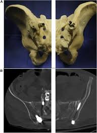
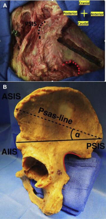
Osteology of the ilium revised: illuminating the clinical relevance

Eur J Trauma Emerg Surg. 2020 Sep 4. doi: 10.1007/s00068-020-01482-2.Online ahead of print.

Alexander von Glinski, Sven Frieler, Emre Yilmaz, Basem Ishak, Ryan Goodmanson, Joe Iwanaga, Thomas A Schildhauer, Jens R Chapman, Rod J Oskouian, Keith Mayo, R Shane Tubbs


Abstract

Background: Several studies on anterior and posterior pelvic ring fixation have identified a fragile monocortical area located at the iliac wing. However, there are no current studies regarding this structure's dimensions and relation to known anatomic structures.

Methods: Eleven human ilia were dissected from 6 specimens. After debulking soft tissue, photoluminescence was used to indicate the fragile area. The size and thickness of the iliac wing were determined and mapped in relation to the anterior superior iliac spine (ASIS) and the posterior superior iliac spine (PSIS).

Results: This photoluminescent unicortical area measured 15.57 cm2 with a mean minimal thickness of 1.37 mm at its thinnest part. Its average diameter was 41.15 mm horizontally and 37.45 mm vertically. In all cases, it was located at the middle third of the ilium with a mean distance of 64.58 mm to the AIIS and 62.73 mm to the PSIS. Trajectory angulation above 4.5° from the PSIS lead to violation of this area.

Conclusion: This study provides useful anatomical information regarding a thin unicortical area at the iliac wing that is relevant to anterior and posterior pelvic ring fixation and the potential complications that can arise from iatrogenic perforation of this area.

Keywords: Ilium osteology; Pelvic anatomy; Sacropelvic fixation; Tricortical iliac graft.R Shane Tubbs


Tyler LawsComment
De novo methicillin-resistant Staphylococcus aureus vs. methicillin-sensitive Staphylococcus aureus infections of the spine

Abstract

Vertebral osteomyelitis (VO) is a severe infection of the vertebral body and the adjacent disc space, where Staphylococcus aureus is most commonly isolated. The objective of this retrospective study was to determine risk factors for and compare outcome differences between de novo methicillin-resistant Staphylococcus aureus (MRSA) VO and methicillin-sensitive Staphylococcus aureus (MSSA) VO. A retrospective cohort study was performed by review of the electronic medical records of 4541 consecutive spine surgery patients. Among these 37 underwent surgical treatment of de novo MRSA and MSSA spinal infections. Patient demographics, pre- and postoperative neurological status (ASIA impairment score), surgical treatment, inflammatory laboratory values, nutritional status, comorbidities, antibiotics, hospital stay, ICU stay, reoperation, readmission, and complications were collected. A minimum follow-up (FU) of 12 months was required. Among the 37 patients with de novo VO, 19 were MRSA and 18 were MSSA. Mean age was 52.4 and 52.9 years in the MRSA and MSSA groups, respectively. Neurological deficits were found in 53% of patients with MRSA infection and in 17% of the patients with MSSA infection, which was statistically significant (p < 0.05). Chronic renal insufficiency and malnutrition were found to be significant risk factors for MRSA VO. Preoperative albumin was significantly lower in the MRSA group (p < 0.05). Patients suffering from spinal infection with chronic renal insufficiency and malnutrition should be watched more carefully for MRSA. The MRSA group did not show a significant difference with regard to final clinical outcome despite more severe presentation.

Keywords: Albumin; MRSA; MSSA; Malnutrition; Spinal infection; Vertebral osteomyelitis.


Tyler LawsComment
Foraminal Ligaments Tether Upper Cervical Nerve Roots: A Potential Cause of Postoperative C5 Palsy

J Brachial Plex Peripher Nerve Inj. 2020 Jul 24;15(1):e9-e15. doi: 10.1055/s-0040-1712982. eCollection 2020 Jan.

Andrew S Jack, Brooks R Osburn, Zane A Tymchak, Wyatt L Ramey, Rod J Oskouian, Robert A Hart, Jens R Chapman, Line G Jacques, R Shane Tubbs


Abstract

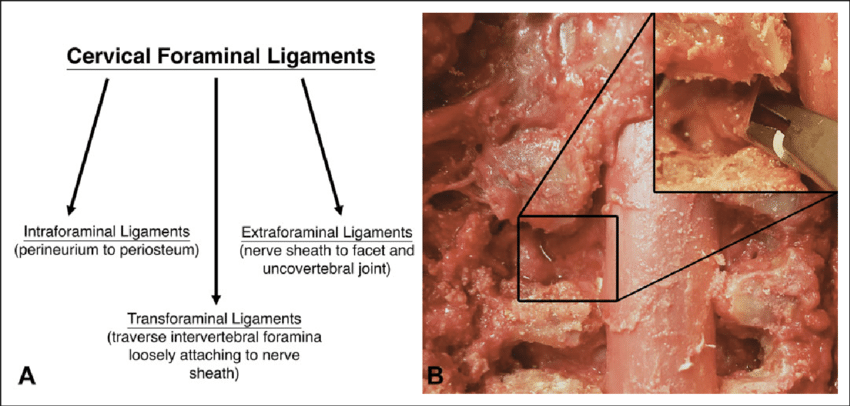
Background Nerve root tethering upon dorsal spinal cord (SC) migration has been proposed as a potential mechanism for postoperative C5 palsy (C5P). To our knowledge, this is the first study to investigate this relationship by anatomically comparing C5-C6 nerve root translation before and after root untethering by cutting the cervical foraminal ligaments (FL). Objective The aim of this study is to determine if C5 root untethering through FL cutting results in increased root translation. Methods Six cadaveric dissections were performed. Nerve roots were exposed via C4-C6 corpectomies and supraclavicular brachial plexus exposure. Pins were inserted into the C5-C6 roots and adjacent foraminal tubercle. Translation was measured as the distance between pins after the SC was dorsally displaced 5 mm before and after FL cutting. Clinical feasibility of FL release was examined by comparing root translation between standard and extended (complete foraminal decompression) foraminotomies. Translation of root levels before and after FL cutting was compared by two-way repeated measures analysis of variance. Statistical significance was set at 0.05. Results Significantly more nerve root translation was observed if the FL was cut versus not-cut, p = 0.001; no difference was seen between levels, p = 0.33. Performing an extended cervical foraminotomy was technically feasible allowing complete FL release and root untethering, whereas a standard foraminotomy did not. Conclusion FL tether upper cervical nerve roots in their foramina; cutting these ligaments untethers the root and increases translation suggesting they could be harmful in the context of C5P. Further investigation is required examining the value of root untethering in the context of C5P.


Tyler LawsComment
Antibiotic-impregnated polymethylmethacrylate strut graft as a treatment of spinal osteomyelitis: case series and description of novel technique.

J Neurosurg Spine. 2020 May 8;1-6. doi: 10.3171/2020.3.SPINE191313.Online ahead of print.

Wyatt L Ramey, Alexander von Glinski, Andrew Jack, Ronen Blecher, Rod J Oskouian, Jens R Chapman


Abstract

Objective: The surgical treatment of osteomyelitis and discitis of the spine often represents a challenging clinical entity for a multitude of reasons, including progression of infection despite debridement, development of spinal deformity and instability, bony destruction, and seeding of hardware. Despite advancement in spinal hardware and implantation techniques, these aforementioned challenges not uncommonly result in treatment failure, especially in instances of heavy disease burden with enough bony endplate destruction as to not allow support of a modern titanium cage implant. While antibiotic-infused polymethylmethacrylate (aPMMA) has been used in orthopedic surgery in joints of the extremities, its use has not been extensively described in the spine literature. Herein, the authors describe for the first time a series of patients treated with a novel surgical technique for the treatment of spinal osteomyelitis and discitis using aPMMA strut grafts with posterior segmental fusion.

Methods: Over the course of 3 years, all patients with spinal osteomyelitis and discitis at a single institution were identified and included in the retrospective cohort if they were surgically treated with spinal fusion and implantation of an aPMMA strut graft at the nidus of infection. Basic demographics, surgical techniques, levels treated, complications, and return to the operating room for removal of the aPMMA strut graft and placement of a traditional cage were examined. The surgical technique consisted of performing a discectomy and/or corpectomy at the level of osteomyelitis and discitis followed by placement of aPMMA impregnated with vancomycin and/or tobramycin into the cavity. Depending on the patient's condition during follow-up and other deciding clinical and radiographic factors, the patient may return to the operating room nonurgently for removal of the PMMA spacer and implantation of a permanent cage with allograft to ultimately promote fusion.

Results: Fifteen patients were identified who were treated with an aPMMA strut graft for spinal osteomyelitis and discitis. Of these, 9 patients returned to the operating room for aPMMA strut graft removal and insertion of a cage with allograft at an average of 19 weeks following the index procedure. The most common infections were methicillin-sensitive Staphylococcus aureus (n = 6) and methicillin-resistant S. aureus (n = 5). There were 13 lumbosacral infections and 1 each of cervical and thoracic infection. Eleven patients were cured of their infection, while 2 had recurrence of their infection; 2 patients were lost to follow-up. Three patients required unplanned return trips to the operating room, two of which were for wound complications, with the third being for recurrent infection.

Conclusions: In cases of severe infection with considerable bony destruction, insertion of an aPMMA strut graft is a novel technique that should be considered in order to provide strong anterior-column support while directly delivering antibiotics to the infection bed. While the active infection is being treated medically, this structural aPMMA support bridges the time it takes for the patient to be converted from a catabolic to an anabolic state, when it is ultimately safe to perform a definitive, curative fusion surgery.


Tyler LawsComment
Facing multidrug-resistant pathogens in periprosthetic joint infections with self-administered outpatient parenteral antimicrobial therapy-A prospective cohort study

J Orthop Res. 2021 Feb;39(2):320-332. doi: 10.1002/jor.24906.Epub 2020 Dec 1.

Sven Frieler, Yannik Hanusrichter, Petri Bellova, Jan Geßmann, Thomas A Schildhauer, Hinnerk Baecker


Abstract

A key factor in the successful management of periprosthetic joint infection (PJI) besides the surgical regime is a consistent antimicrobial therapy. Recently, oral versus intravenous (IV) antibiotics for bone and joint infection trial demonstrated the noninferiority of oral antimicrobial therapy compared to IV, implying that an early transition to oral administration is reasonable. It is likely that the international consensus meeting of musculoskeletal Infections and the European Bone and Joint Infection Society will consider these findings. However, rising levels of antimicrobial resistance are challenging and recommendations for dealing with multidrug-resistant (MDR) pathogens resistant to oral antibiotics are lacking. This study focuses on establishing guidance towards their management in PJI. From December 2015 to June 2019, patients with MDR pathogens were included in a single-center prospective cohort study and treated with self-administered outpatient parenteral antimicrobial therapy (S-OPAT) based on a two-stage revision strategy. Demographics, pathogens, antimicrobial agents, and outcomes were recorded. A total of 1738 outpatient days in 26 patients were analyzed. The incidence of pathogens resistant to oral antibiotics in PJI was 4%, most frequently encountered were staphylococcus epidermidis. The Kaplan-Meier-estimated infection-free survival after 3 years was 90% (95% confidence interval, 84.6%-95.5%). We recorded adverse events in 6 of 54 (11%) S-OPAT episodes (3.45/1000 S-OPAT days). (i) S-OPAT in two-stage revision arthroplasty to counter increasing numbers of MDR pathogens resistant to oral agents can achieve a high infection eradication rate and (ii) should therefore be taken into account at the next society's consensus treatment updates.

Tyler LawsComment
Prevention of Wrong-level Surgery in the Thoracic Spine: Preoperative Computer Tomography Fluoroscopy-guided Percutaneous Gold Fiducial Marker Placement in 57 Patients.

Spine (Phila Pa 1976). 2020 Dec 15;45(24):1720-1724. doi: 10.1097/BRS.0000000000003691.

Basem Ishak, Amir Abdul-Jabbar, Tamir Tawfik, Emre Yilmaz, Alexander von Glinski, Andreas Unterberg, Rod Oskouian, Jens R Chapman


Abstract

Study design: Retrospective review.

Objective: The aim of this study was to evaluate the feasibility, safety,s and complications of computer tomography (CT) fluoroscopy-guided percutaneous transpedicular gold fiducial marker insertion to reduce incidence of wrong-level surgery in the thoracic spine.

Summary of background data: Intraoperative localization of the correct thoracic level can be challenging and time-consuming, especially in obese patients and patients with anatomical variations. In the literature there are very few studies containing low numbers of patients which assessed CT or CT fluoroscopy-guided fiducial marker placement of the thoracic spine. Description of this technique has been similarly scarce.

Methods: All patients who underwent percutaneous CT fluoroscopy-guided gold fiducial marker placement of the thoracic spine were retrospectively reviewed. Indications for surgery included degenerative disc disease, infection, spinal metastasis, and intra- and extradural tumors. Gold fiducial markers were placed using a percutaneous CT fluoroscopy-guided transpedicular approach with local anesthesia. In addition, sex, age, body mass index (BMI), thoracic level, related pathology, and procedure-related complications were also recorded.

Results: A total of 57 patients (24 females, 33 males) were included. Mean age was 58.6 ± 15.5 years. No complications during CT fluoroscopy-guided gold fiducial marker placement were recorded. Intraoperative localization was successful in all patients. Mean BMI was 32.98 kg/m (range, 18.63-56.03 kg/m), and 63% of patients were obese (>30 kg/m). T7 (n = 11) was the most often marked vertebral body, followed by T10 (n = 10) and T6 (n = 7). The most cranial and most caudal levels marked were T2 and T12, respectively.

Conclusion: Preoperative CT fluoroscopy-guided percutaneous gold fiducial marker placement is safe, feasible, and accurate. The resulting facilitated localization of the intended thoracic level of surgery can reduce the length of surgery and prevent wrong-level surgery. Further studies are needed to evaluate in the effect on exposure to radiation and quantify the difference in operating room time.


Tyler LawsComment
What Are Risk Factors for an Ileus After Posterior Spine Surgery?-A Case Control Study

Global Spine J. 2021 Jan 12;2192568220981971. doi: 10.1177/2192568220981971.Online ahead of print.

Emre Yilmaz, Eric Benca, Akil P Patel, Sarah Hopkins, Ronen Blecher, Amir Abdul-Jabbar, Thomas M O'Lynnger, Rod J Oskouian, Daniel C Norvell, Jens Chapman


Abstract

Study design: Case-Control Study.

Objective: The purpose of this retrospective study is to evaluate risk factors for developing a postoperative ileus after posterior spine surgery.

Methods: Patient charts, including radiographs were evaluated retrospectively. Diagnosis of an ileus was confirmed radiographically by a CT scan in all cases. The control group was retrieved by selecting a random sample of patients undergoing posterior spine surgery who did not develop bowel dysfunction postoperatively.

Results: A total of 40 patients had a postoperative ileus. The control group consisted of 80 patients. Both groups did not differ significantly in age, gender, BMI, tobacco use, comorbidities or status of previous abdominal surgery. Significant differences between the 2 groups was the length of stay (5.9 vs. 11.2; p = 0.001), surgery in the lumbar spine (47.5% vs. 87.5%; p < 0.001) and major spine surgery involving > 3 levels (35.0% vs. 57.5%; p = 0.019). Patients who suffered from an ileus were more likely to be treated in ICU (23.8% vs. 37.5%; p = 0.115), being re-admitted (0.0% vs 5.0%; p = 0.044) and having a delayed discharge (32.5% vs. 57.5%; p = 0.009). Multivariable analysis demonstrated that lumbar spine surgery compared to thoracic and/or cervical spine surgery (p = 0.00, OR 8.7 CI 2.9-25.4) and major spine surgery involving > 3 levels (p = 0.012; OR 3.0, CI 1.3-7.2) are associated with developing an ileus postoperatively.

Conclusion: Surgeries of the lumbar spine as well as those involving > 3 levels are associated with developing a postoperative ileus. Further studies are needed to expand on possible risk factors and to better understand the mechanism underlying postoperative ileus in spine surgery patients.

Keywords: complications; lumbar spine surgery; posterior spine surgery; postoperative ileus; spine surgery.


Tyler LawsComment
Quality and Clinical Care Development in Spine Surgery-Connecting the Dots: An Expanded Clinical Narrative

Global Spine J. 2020 Jan;10(1 Suppl):10S-16S. doi: 10.1177/2192568219871248.Epub 2020 Jan 6.

Sarah Hopkins, Polly Brune, Jens R Chapman, Marc Horton, Rod Oskouian, Akshal Patel, Marc D Moisi


Abstract

Our health care system is an evidenced-based quality-centric environment. Pursuit of quality is a process that encompasses knowledge development and care advancements through collaboration and expertise. Depicted here is the foundational knowledge, process, and contributions that hallmark successful clinical quality programs. Beginning with methodology, followed by process and form, we create the foundational knowledge and exemplars demonstrating framework and continuum of process in pursuit and attainment of successful clinical quality and care development for patients. Although our protocol has been devised for complex spine care, this could be implemented across all health care specialties to provide individualized and high-quality care for all current and future patients, all while creating a culture of accountability for physicians.


Tyler LawsComment
Conversion From Knee Arthrodesis Back to Arthroplasty: A Particular Challenge in Combination With Fungal Periprosthetic Joint Infection

Arthroplast Today. 2020 Dec 5;6(4):1038-1044. doi: 10.1016/j.artd.2020.10.007. eCollection 2020 Dec.

Sven Frieler, Emre Yilmaz, Ryan Goodmanson, Yannik Hanusrichter, Thomas A Schildhauer, Hinnerk Baecker


Abstract

A 58-year-old female treated at an outside facility with knee arthrodesis due to persistent periprosthetic joint infection fulfilled all prerequisites for a conversion back to arthroplasty, as part of a 2-stage revision. Owing to the detection of Candida parapsilosis, the treatment concept was converted to a three-stage procedure. A scheduled spacer exchange with additional amphotericin B-loaded polymethylmethacrylate was conducted as an intermediate revision before reimplantation. Conversion in the setting of fungal periprosthetic joint infection presents a challenge, and successful treatment hinges on the use of proper antifungal and antimicrobial protocols, advanced surgical techniques, and a multidisciplinary team approach. At the 3-year follow-up, successful infection eradication as measured by the Delphi-based consensus definition was achieved with a range of motion of 0°-100°.

Keywords: Arthroplasty; Conversion; Fungal periprosthetic joint infection; Knee arthrodesis; PJI; TKA; Three-stage revision; fPJI.

Tyler LawsComment