Risk Factors Associated with 90-day Readmissions Following Occipitocervical Fusion- A Nationwide Readmissions Database Study

World Neurosurg. 2020 Dec 12;S1878-8750(20)32597-3. doi: 10.1016/j.wneu.2020.12.031.Online ahead of print.

Christopher Elia, Ariel Takayanagi, Varun Arvind, Ryan Goodmanson, Alexander von Glinski, Clifford Pierre, Jeanju Sung, Bilal Qutteineh, Edward Jung, Jens Chapman, Rod Oskouian


Background

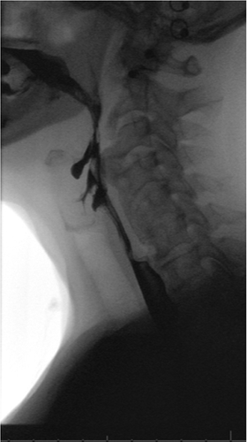
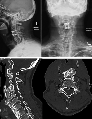
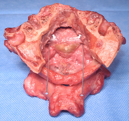
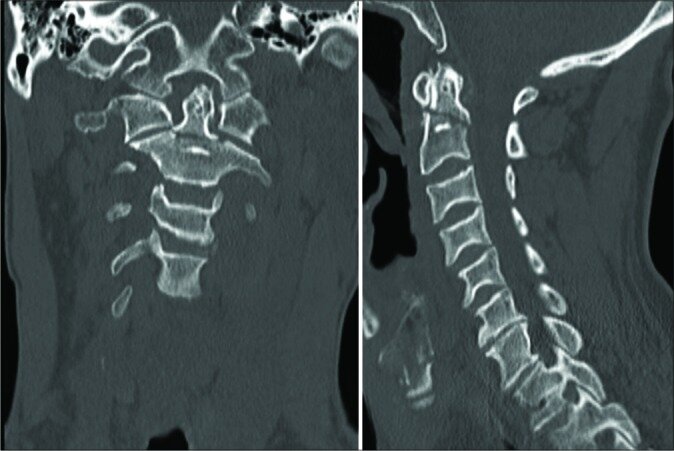
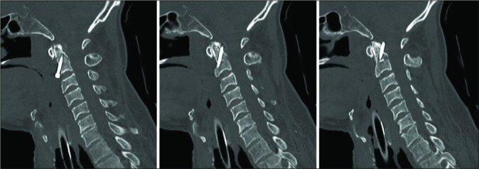
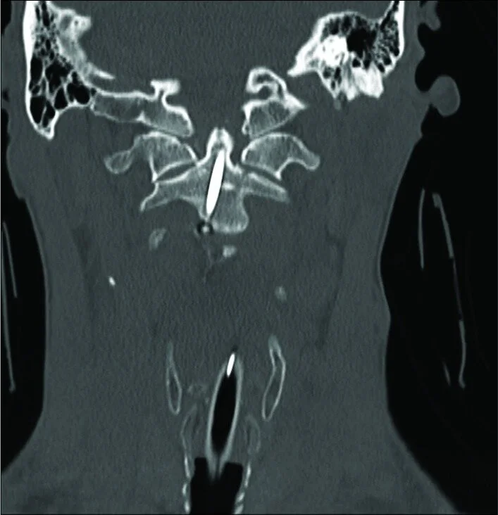
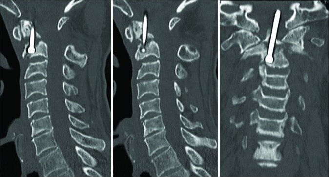
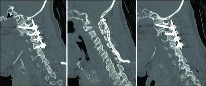
Occipitocervical fusion (OCF) procedures are increasing due to an aging population and the prevalence of trauma, rheumatoid arthritis, and tumors. Reoperation rates and readmission risk factors for cervical fusions have been established, but in relation to OCF they have not been explored. This study investigates the patterns of readmissions and complications following OCF using a national database.

Methods

The 2016 U.S. Nationwide Readmissions Database was used for sample collection. Adults (>18 years) who underwent OCF were identified using the 2016 ICD-10 coding system, and we examined the readmission rates (30-day and 90-day) and reoperation rates.

Results

Between January and September 2016, a total of 477 patients underwent OCF; the 30-day and 90-day readmission rates were 10.4% and 22.4%, respectively. The 90-day reoperation rate related to the index surgery was 5.7%. Mean age (68.58 years) was significantly greater in the readmitted group versus nonreadmitted group (61.76 years) (P < 0.001). The readmitted group had a significantly higher Charlson Comorbidity Index and Elixhauser Comorbidity Index (5.00 and 2.41, respectively) than the nonreadmitted group (3.25 and 1.15, respectively; P < 0.001). Nonelective OCF showed a higher readmission rate (29.18%) versus elective OCF (12.23%) (P < 0.001). Medicare and Medicaid patients showed the highest rates of readmission (27.27% and 20.41%, respectively). Readmitted patients had higher total health care costs.

Conclusions

Nonelective OCF was found to have a readmission rate of almost 2½× that of elective OCF. Understanding risk factors associated with OCF will help with operative planning and patient optimization.

Key words

Cervical fusion, Cervical spine surgery, Nationwide Readmissions Database, Occipitocervical fusion, Readmission rates, Rheumatoid arthritis

Tyler LawsComment
Facing multidrug-resistant pathogens in periprosthetic joint infections with self-administered outpatient parenteral antimicrobial therapy-A prospective cohort study

J Orthop Res. 2020 Nov 11. doi: 10.1002/jor.24906.Online ahead of print.

Sven Frieler, Yannik Hanusrichter, Petri Bellova, Jan Geßmann, Thomas A Schildhauer, Hinnerk Baecker


Abstract

A key factor in the successful management of periprosthetic joint infection (PJI) besides the surgical regime is a consistent antimicrobial therapy. Recently, oral versus intravenous (IV) antibiotics for bone and joint infection trial demonstrated the noninferiority of oral antimicrobial therapy compared to IV, implying that an early transition to oral administration is reasonable. It is likely that the international consensus meeting of musculoskeletal Infections and the European Bone and Joint Infection Society will consider these findings. However, rising levels of antimicrobial resistance are challenging and recommendations for dealing with multidrug-resistant (MDR) pathogens resistant to oral antibiotics are lacking. This study focuses on establishing guidance towards their management in PJI. From December 2015 to June 2019, patients with MDR pathogens were included in a single-center prospective cohort study and treated with self-administered outpatient parenteral antimicrobial therapy (S-OPAT) based on a two-stage revision strategy. Demographics, pathogens, antimicrobial agents, and outcomes were recorded. A total of 1738 outpatient days in 26 patients were analyzed. The incidence of pathogens resistant to oral antibiotics in PJI was 4%, most frequently encountered were staphylococcus epidermidis. The Kaplan-Meier-estimated infection-free survival after 3 years was 90% (95% confidence interval, 84.6%-95.5%). We recorded adverse events in 6 of 54 (11%) S-OPAT episodes (3.45/1000 S-OPAT days). (i) S-OPAT in two-stage revision arthroplasty to counter increasing numbers of MDR pathogens resistant to oral agents can achieve a high infection eradication rate and (ii) should therefore be taken into account at the next society's consensus treatment updates.

Keywords: DTT; MDR pathogen; OPAT; PJI; two-stage revision.

Tyler LawsComment
Osteology of the ilium revised: illuminating the clinical relevance

Eur J Trauma Emerg Surg. 2020 Sep 4. doi: 10.1007/s00068-020-01482-2.Online ahead of print.

Alexander von Glinski, Sven Frieler, Emre Yilmaz, Basem Ishak, Ryan Goodmanson, Joe Iwanaga, Thomas A Schildhauer, Jens R Chapman, Rod J Oskouian, Keith Mayo, R Shane Tubbs


Abstract

Background: Several studies on anterior and posterior pelvic ring fixation have identified a fragile monocortical area located at the iliac wing. However, there are no current studies regarding this structure's dimensions and relation to known anatomic structures.

Methods: Eleven human ilia were dissected from 6 specimens. After debulking soft tissue, photoluminescence was used to indicate the fragile area. The size and thickness of the iliac wing were determined and mapped in relation to the anterior superior iliac spine (ASIS) and the posterior superior iliac spine (PSIS).

Results: This photoluminescent unicortical area measured 15.57 cm2 with a mean minimal thickness of 1.37 mm at its thinnest part. Its average diameter was 41.15 mm horizontally and 37.45 mm vertically. In all cases, it was located at the middle third of the ilium with a mean distance of 64.58 mm to the AIIS and 62.73 mm to the PSIS. Trajectory angulation above 4.5° from the PSIS lead to violation of this area.

Conclusion: This study provides useful anatomical information regarding a thin unicortical area at the iliac wing that is relevant to anterior and posterior pelvic ring fixation and the potential complications that can arise from iatrogenic perforation of this area.

Keywords: Ilium osteology; Pelvic anatomy; Sacropelvic fixation; Tricortical iliac graft.

Tyler Laws Comment
Extreme Lateral Interbody Fusion for Thoracic and Thoracolumbar Disease: The Diaphragm Dilemma

Global Spine J. 2020 Mar 27;2192568220914883. doi: 10.1177/2192568220914883. Online ahead of print.

Alexander Von GlinskiChristopher J EliaAriel TakayanagiEmre YilmazBasem IshakJoe DettoriBenjamin A SchellErik HaymanClifford PierreJens R ChapmanRod J Oskouian


Abstract

Study design: Retrospective cohort study.

Objective: Complication profiles for lateral approaches to the spine are well established. However, the influence of level of surgery on complication rates and subtypes are less well established. To determine risk factors for complications as determined by level and surgery type in patients undergoing a lateral (retroperitoneal or retropleural approach) to the thoracolumbar spine.

Methods: All adult patients undergoing a lateral thoracolumbar fusion with or without posterior instrumentation performed at a single institution were identified. Primary outcomes assessed were presence of complication, complication subtype, and need for reoperation. The primary independent variables were spinal level (thoracic, thoracolumbar, or lumbar) and type of surgery (discectomy or corpectomy). Categorical outcomes were compared using chi-square test. Unadjusted and adjusted odds ratios for corpectomy status were calculated to determine risk of complication by level. P < .05 was considered statistically significant.

Results: A total of 165 patients aged 18 to 75 years were identified as having undergone a lateral fusion. Complication rates were 28.6%, 36.4%, and 11% for thoracic, thoracolumbar, and lumbar lateral approach fusions, respectively. Under univariate analysis, patients undergoing lateral approach in the thoracic spine group had significantly higher rates of postoperative complications than those in the lumbar group (P = .005). After adjusting for corpectomy status, there was no difference in complication rates.

Conclusions: Lateral (retroperitoneal or retropleural) approaches to the thoracic and thoracolumbar spine may be used with complication rates comparable to well-established lumbar approaches. Extent of surgery (corpectomy vs discectomy) rather than level of surgery may represent the primary driver of complications.

Keywords: complication; corpectomy; discectomy; lateral approach; neurologic deficit; pneumothorax.

Tyler LawsComment
Space-Occupying Lesions of the Retropharyngeal Space: An Anatomical Study With Application to Postoperative Retropharyngeal Hematomas

Global Spine. J 2020 May 13;2192568220922192. doi: 10.1177/2192568220922192. Online ahead of print.

Alexander von GlinskiChristopher EliaEmre YilmazSven FrielerBasem IshakMahindra Kumar AnandJoe IwanagaAmir Abdul-JabbarRod J OskouianR Shane TubbsJens R Chapman


Abstract

Study design: Cadaver study.

Objective: The retropharyngeal space's (RPS's) clinical relevance is apparent in anterior cervical spine surgery with respect to postoperative hematoma, which can cause life-threatening airway obstruction. This cadaver study aims to establish guidance toward a better understanding of the tolerance of the RPS to accommodate fluid accumulation.

Methods: Five fresh-frozen cadavers were dissected in the supine position. A digital manometer and a 20 Fr Foley catheter were inserted into the RPS via an anterolateral approach. While inflating the Foley catheter, the position of the esophagus/trachea was documented using fluoroscopy, and the retropharyngeal pressure was measured. We quantified the volume required to deviate the esophagus/trachea >1 cm from its original position using fluoroscopy. We also recorded the volume required to cause a visible change to the normal neck contour.

Results: A mean volume of 12.5 mL (mean pressure 1.50 mm Hg) was needed to cause >1 cm of esophageal deviation. Tracheal deviation was encountered at a mean volume of 20.0 mL (mean pressure of 2.39 mm Hg). External visible clinical neck contour changes were apparent at a mean volume of 39 mL.

Conclusion: A relatively small volume of fluid in the RPS can cause the esophagus/trachea to radiographically deviate. The esophagus is the structure in the RPS to be most influenced by mass effect. The mean volume of fluid required to cause clinically identifiable changes to the normal neck contour was nearly double the volume required to cause 1 cm of esophageal/tracheal deviation in a cadaver model.

Keywords: airway complications; anatomical study; dysphagia; retropharyngeal hematoma; retropharyngeal space; trachea deviation.

Tyler LawsComment
Surgical Treatment of Ossifications of the Cervical Anterior Longitudinal Ligament: A Retrospective Cohort Study

Global Spine J. 2020 May 19;2192568220922195. doi: 10.1177/2192568220922195. Online ahead of print.

Alexander von GlinskiAriel TakayanagiChristopher EliaBasem IshakMishan ListmannClifford A PierreRonen BlecherErik HaymanJens R ChapmanRod J Oskouian


Abstract

Study design: Retrospective cohort study.

Objectives: The study aims to evaluate anterior cervical discectomy and fusion (ACDF) in the treatment of patients with ossification of the anterior longitudinal ligament (OALL).

Methods: We retrospectively reviewed cases performed at our institution between January 2015 and December 2018; adult (age ≥18 years) patients who underwent anterior cervical decompression and fusion in the presence of dysphagia and OALL. Ten patients (9 male, 1 female, mean age 64.4 years) with OALL who underwent ACDF were included. Charts were reviewed for demographics and comorbidities. Primary outcomes assessed were intra- and postoperative complications. Secondary outcomes were fusion rates, instrumentation failure, postsurgical instability/deformity, and readmission rates.

Results: The average duration of symptoms prior to surgery was 12.3 months. All patients presented with dysphagia (mean Bazaz score 2.0). The average number of levels with OALL was 4.7 (±1.67). All patients underwent ACDF and 3 patients underwent additional posterior cervical fusion for kyphotic deformity correction or when extensive laminectomy was required. We did not encounter any intraoperative complications. Eight patients (72%) had solid fusion demonstrated on the lateral x-rays and no evidence of progressive kyphotic deformity. We did not encounter any instrumentation failure or loosening. Two patients developed recurrence of dysphagia (Bazaz scores 2 and 3 respectively).

Conclusion: ACDF for OALL with dysphagia and concomitant myelopathy in our small series of 10 patients demonstrate good fusion and clinical outcomes. Larger studies will be necessary to determine the optimal treatment for patients with dysphagia due to OALL.

Keywords: ACDF; OALL; OPLL; anterior osteophytes; diffuse idiopathic skeletal hyperostosis (DISH); dysphagia.

Tyler LawsComment
The new onset of dysphagia four years after anterior cervical discectomy and fusion: Case report and literature review

Surg Neurol Int . 2020 Feb 28;11:32. doi: 10.25259/SNI_46_2020. eCollection 2020.

Darius AnsariHalle E K BurleyAlexander von GlinskiChristopher EliaJens R ChapmanRod J Oskouian


Abstract

Background: Dysphagia is a common complication immediately following anterior cervical spine surgery. However, its onset more than 1-year postoperatively is rare.

Case description: A 45-year-old male initially underwent a C3-4 and C5-6 anterior cervical discectomy and fusion (ACDF). At age 49, 4 years later, he presented with worsening dysphagia accompanied by neck and right upper extremity pain. Radiographs demonstrated an extruded left C3 screw, which had migrated into the prevertebral soft tissues at the C4-C5 level; there was also loosening of the right C3 screw. The subsequent barium swallow study revealed that the screw was embedded in the pharyngeal wall. The patient required a two-stage operation; first, to remove the anterior instrumentation, and second, to perform a posterior instrumented C2-T2 fusion.

Conclusion: A barium swallow study and other dynamic imaging are a valuable component of the diagnostic workup and therapeutic intervention to evaluate the delayed onset dysphagia following an ACDF.

Keywords: Anterior cervical discectomy and fusion; Complications; Delayed onset dysphagia; Hardware failure.

Copyright: © 2020 Surgical Neurology International.


Tyler LawsComment
Emerging Insight in the Use of an Active Post Discharge Surveillance Program in Spine Surgery: A Retrospective Pilot Study

World Neurosurg . 2020 Jul;139:e237-e244. doi: 10.1016/j.wneu.2020.03.185. Epub 2020 Apr 14.

Alexander von GlinskBasem IshakChristopher J EliaRyan GoodmansonClifford PierreDaniel C NorvellDarius AnsariPolly BruneRod J OskouianJens R Chapman


Abstract

Background: With smartphones being present in everyday life, we have witnessed an increasing use of applications designed for mobile communication devices that are aimed at facilitating patient engagement in different medical arenas. Such applications are meant to improve communications with patients and ultimately improve patient care. The aim of this study was to report on our early experiences using Active Post Discharge Surveillance (APDS) relative to invasiveness of the spine surgery and patient age and gender.

Methods: A retrospective chart review was performed including all patients who volunteered to use application-based APDS between September 1, 2017, and September 30, 2018. The primary outcome was the number of APDS uses. Secondary outcomes were inquiries that led to a change of treatment or induced a readmission and patient satisfaction. Regression analysis was performed regarding the influence of invasiveness, age, and gender on the incidence of APDS use.

Results: The average number of individual APDS communications was 3.6 with no difference between degrees of severity of invasive surgery, age, or gender. APDS inquiries induced unexpected readmissions in 4 patients (66.6% of all readmissions) and postoperative treatment regimen changes in 4 other patients. Thirty-three patients (86.8%) reported being satisfied with APDS usage.

Conclusions: This is the first study to investigate use of interactive APDS in patients undergoing elective spine surgery. Our data suggest that patient age, gender, or invasiveness of surgery is not associated with the usage of APDS.

Keywords: Active Post Discharge Surveillance; Apps; Mobile applications; Smartphone.

Copyright © 2020 Elsevier Inc. All rights reserved.

Tyler LawsComment
Case Report of a Pelvic Crossed Fused Renal Ectopic Kidney

Kurume Med J . 2020 Jul 1;66(1):55-58. doi: 10.2739/kurumemedj.MS661004. Epub 2020 Apr 24.

Emily SimondsJoe IwanagaShogo KikutaMaia SchumacherGraham DupontJuan AltafullaEmre YilmazRod J OskouianR Shane Tubbs


Abstract

Herein we present a case of a horseshoe kidney with crossed fused renal ectopia. Both of these pathologies are congenital anomalies; however, to date, there are few cases that present with both. In this case, discovered during routine dissection, the fused kidney was mostly left-sided and very low in the pelvis. No renal artery arose from the right wall of the abdominal aorta, and the right renal vein drained into the lower part of the inferior vena cava (IVC) where the right and left common iliac veins joined. It is essential for clinicians and surgeons to understand these types of congenital anomalies, as they could impact patient care.

Keywords: Ectopic kidney; cadaver, anatomy; cross fused renal ectopia; horseshoe kidney; inferior vena cava; renal artery.


Tyler LawsComment
Update on the Biomechanics of the Craniocervical Junction, Part II: Alar Ligament

Global Spine J . 2020 Jul 21;2192568220941452. doi: 10.1177/2192568220941452. Online ahead of print.

Basem IshakAlexander von GlinskiGraham Dupont , Stefan LachkarEmre YilmazJoe IwanagaAndreas UnterbergRod OskouianR Shane TubbsJens R Chapman


Abstract

Study design: In vitro biomechanical study.

Objective: The strength of the alar ligament has been described inconsistently, possibly because of the nonphysiological biomechanical testing models, and the inability to test the ligament with both attachments simultaneously. The purpose of this biomechanical model was to reevaluate the alar ligament's tensile strength with both bony attachments, while also keeping the transverse ligament intact, all in a more physiological biomechanical model that mimics the mechanism of traumatic injury closely.

Methods: Eleven fresh-frozen occipito-atlanto-axial (C0-C1-C2) specimens were harvested from individuals whose mean age at death was 77.4 years (range 46-97 years). Only the alar and transverse ligaments were preserved, and the bony C0-C1-C2 complex was left intact. Axial tension was exerted on the dens to displace it posteriorly, while the occipito-axial complex was fixed anteriorly. A device that applies controlled increasing force was used to test the tensile strength (M2-200, Mark-10 Corporation).

Results: The mean force required for the alar ligament to fail was 394 ± 52 N (range 317-503 N). However, both the right and left alar ligaments ruptured simultaneously in 10 specimens. The ligament failed most often at the dens (n = 10), followed by occipital condyle rupture (n = 1). The transverse ligament remained intact in all specimens.

Conclusions: When both the right and left alar ligament were included, the total alar ligament failure occurred at an average force of 394 N. The alar ligament failed before the transverse ligament.

Keywords: alar ligament; biomechanical study; cadaveric study; craniocervical junction; failure force.


Tyler LawsComment
Postoperative intracranial migration of a C2 odontoid screw: A case report and literature review.

Surg Neurol Int. 2019 Sep 10;10:173. doi: 10.25259/SNI_245_2019. eCollection 2019.

Ankush ChandraSeong-Jin MoonBlake WalkerEmre YilmazMarc MoisiRobert Johnson


Abstract

Background: Intracranial migration of odontoid screws is a rare but serious complication of anterior odontoid screw fixation not often reported in literature by neurosurgeons. Here, we describe the second case in literature of intracranial migration of an odontoid screw.

Case description: A 64-year-old neurologically intact patient with a type II odontoid fracture secondary to trauma underwent anterior odontoid screw fixation without any intraoperative complications. He tolerated the procedure well, and postoperative imaging demonstrated near anatomic correction of the fracture with satisfactory placement of the lag screw. Unfortunately, the patient was subsequently lost to follow up and he presented 7 months later for a routine outpatient computed tomography (CT) of the cervical spine, which demonstrated upward migration of the screw into the intracranial cavity abutting the medulla, with CT angiography of the neck also confirming the screw lying between the two vertebral arteries. Magnetic resonance imaging of the cervical spine also demonstrated the odontoid screw lying within close proximity to the ventral cervicomedullary junction, marginating the left vertebral artery. Subsequently, the patient was managed with removal of the odontoid screw and posterior cervical arthrodesis and instrumented fusion.

Conclusion: Our case demonstrates the rare but serious complication of intracranial odontoid screw migration, which we bring to the attention of the neurosurgical community. The recognition of risk factors for this complication and optimized management of this rare occurrence is important for surgeons to recognize.

Keywords: Anterior odontoid screw fixation; C2 odontoid screw; Postoperative intracranial migration.

Copyright: © 2019 Surgical Neurology International.

Tyler LawsComment
Tulip-Screw Head Disjunction from Posterior C2 Fracture Fixation Instrumentation.

Case Rep Orthop. 2020 Feb 24;2020:5824383. doi: 10.1155/2020/5824383. eCollection 2020.

Burley HEKAnsari DSvon Glinski AGoodmanson RSchell BChapman JROskouian RJ.


Abstract

This report presents an unusual case of instrumentation failure after posterior fixation of a C2 fracture and reviews currently available treatment alternatives. The patient, a 53-year-old female, initially presented to the emergency department at an outside facility with acute alcohol intoxication and acute neck pain following a fall from a ladder. CT demonstrated bilateral C2 pars fractures and unstable posteroinferior displacement of the posterior elements. She underwent an emergent C2 open-reduction internal fixation (ORIF) at the outside facility with 3.5 mm polyaxial synapse pedicle screws (DePuy Synthes, Switzerland). There were no known complications and the patient was discharged. Two years after the index operation, cervical CT scan at a different facility revealed that although the fracture was fully healed, bilateral tulip caps had detached from the pedicle screw heads at C2. All implants were removed without postoperative complications. Industry review of alternate lag screws approved for the cervical spine demonstrated that there is not currently an ideal implant for fixation of C2 fractures without fusion. Cannulated trauma screws, which are low profile and would have avoided the instrumentation failure seen here, are not currently FDA approved for the cervical spine.

Copyright © 2020 Halle E. K. Burley et al.

Tyler LawsComment
The Neural Sulcus of the Cervical Vertebrae: A Review of Its Anatomy and Surgical Perspectives.

Cureus. 2020 Jan 18;12(1):e6693. doi: 10.7759/cureus.6693.

Joshi NKlinger NHalalmeh DRTubbs RSMoisi MD.


Abstract

The neural sulcus is a bony channel that spans the transverse process in the subaxial cervical spine. It is located between the anterior and posterior tubercles on either side of the transverse foramen, housing the spinal nerve as it passes through the intervertebral foramina. Although numerous studies have evaluated the anatomy of the cervical spine, very little data on detailed anatomy of the neural sulcus and its implication in cervical spine surgery exist. Here, we review the anatomy of the neural sulcus and surgical considerations. The neural sulcus has important surgical implications, and knowledge of its anatomy is important in considering and planning posterior cervical segmented instrumentation. This increases the ability of the neurosurgeon to choose the best suitable surgical approach to the subaxial cervical spine, allowing good outcomes for the patient.

Copyright © 2020, Joshi et al.

KEYWORDS:

anatomy; cervical; neural sulcus; posterior; screw; surgical

Reattachment of the Multifidus Tendon in Lumbar Surgery to Decrease Postoperative Back Pain: A Technical Note.

Cureus. 2019 Dec 12;11(12):e6366. doi: 10.7759/cureus.6366.

Klinger NYilmaz EHalalmeh DRTubbs RSMoisi MD.


Abstract

The posterior midline approach to the lumbar spine requires significant manipulation of the paraspinal muscles. Muscle detachment and retraction results in iatrogenic damage such as crush injury, devascularization, and denervation, all of which have been associated with postoperative pain. The muscle most directly affected by the posterior approach is the lumbar multifidus (LM), the largest and most medial of the deep lumbar paraspinal muscles. The effects of the posterior approach on the integrity of the LM is concerning, as multiple studies have demonstrated that intraoperative injuries sustained by the LM lead to postoperative muscle atrophy and potentially worsening low back pain. Given the inevitability of intraoperative paraspinal muscle manipulation when using the posterior approach, this technical note describes methods by which surgeons may minimize LM tissue disruption and restore the anatomical position of the LM to ultimately expedite recovery, minimize postoperative pain, and improve patient satisfaction.

Copyright © 2019, Klinger et al.

KEYWORDS:

back pain; lumbar surgery; multifidus muscle; postoperative; reattachment

Anatomical and Biomechanical Study of the Lumbar Interspinous Ligament.

Asian J Neurosurg. 2019 Nov 25;14(4):1203-1206. doi: 10.4103/ajns.AJNS_87_19. eCollection 2019 Oct-Dec.

Iwanaga JSimonds EYilmaz ESchumacher MPatel MTubbs RS.


Abstract

OBJECTIVE:

The lumbar interspinous ligaments (ISLs) are thin and short fibers connecting adjacent spinous processes. However, their morphology is variably described and their biomechanics are not well understood. Therefore, the purpose of this study was to assess the anatomy and biomechanics of the lumbar ISL.

MATERIALS AND METHODS:

Five fresh frozen cadaveric specimens were dissected posteriorly to reveal and study the lumbar ISL. Measurements of the ligaments included the anterior vertical height (length A), the posterior vertical height (length P), and the length (length H) at each lumbar level. Next, 17 lumbar vertebral levels from 6 cadaveric specimens were used for tensile strength testing. The ISLs were subjected to vertically controlled increasing manual tension. The force necessary to disrupt the ISL was recorded.

RESULTS:

All the ISLs ran horizontally in an anterior-posterior direction with a slight curve. The average of length A, length P, and length H on the right sides was 9.82, 9.57, and 20.12 mm, respectively. The average of length A, length P, and length H on the left sides was 11.56, 12.01, and 21.42 mm, respectively. The mean tensile strength of the ISL was 162.33 (N) at L1/2, 85.67 (N) at L2/3, and 79 (N) at L3/4. There was a significant difference in the tensile force between L1/2 and L2/3 and L1/2 and L3/4 (P < 0.05). The ligaments became weaker with a descent along the lumbar levels.

CONCLUSION:

The results of this study might help surgeons understand pathology/trauma of the lumbar vertebral region.

Copyright: © 2019 Asian Journal of Neurosurgery.

KEYWORDS:

Biomechanics; cadaver; lumbar interspinous ligaments; spine; tensile strength

Ansa cervicalis: a comprehensive review of its anatomy, variations, pathology, and surgical applications.

Anat Cell Biol. 2019 Sep;52(3):221-225. doi: 10.5115/acb.19.041. Epub 2019 Aug 26.

Kikuta SJenkins SKusukawa JIwanaga JLoukas MTubbs RS.


Abstract

The ansa cervicalis is a neural loop in the neck formed by connecting the superior root from the cervical spinal nerves (C1-2) and the inferior root descending from C2-C3. It has various anatomical variations and can be an important acknowledgment in specific operations of the neck region. This is a review the anatomy, variations, pathology and clinical applications of the ansa cervicalis.

Copyright © 2019. Anatomy & Cell Biology.

KEYWORDS:

Anatomy; Ansa cervicalis; Cervical spinal nerve; Hypoglossal nerve; Neck; Surgery

Postoperative intracranial migration of a C2 odontoid screw: A case report and literature review.

Surg Neurol Int. 2019 Sep 10;10:173. doi: 10.25259/SNI_245_2019. eCollection 2019.

Chandra AMoon SJWalker BYilmaz EMoisi MJohnson R.


Abstract

BACKGROUND:

Intracranial migration of odontoid screws is a rare but serious complication of anterior odontoid screw fixation not often reported in literature by neurosurgeons. Here, we describe the second case in literature of intracranial migration of an odontoid screw.

CASE DESCRIPTION:

A 64-year-old neurologically intact patient with a type II odontoid fracture secondary to trauma underwent anterior odontoid screw fixation without any intraoperative complications. He tolerated the procedure well, and postoperative imaging demonstrated near anatomic correction of the fracture with satisfactory placement of the lag screw. Unfortunately, the patient was subsequently lost to follow up and he presented 7 months later for a routine outpatient computed tomography (CT) of the cervical spine, which demonstrated upward migration of the screw into the intracranial cavity abutting the medulla, with CT angiography of the neck also confirming the screw lying between the two vertebral arteries. Magnetic resonance imaging of the cervical spine also demonstrated the odontoid screw lying within close proximity to the ventral cervicomedullary junction, marginating the left vertebral artery. Subsequently, the patient was managed with removal of the odontoid screw and posterior cervical arthrodesis and instrumented fusion.

CONCLUSION:

Our case demonstrates the rare but serious complication of intracranial odontoid screw migration, which we bring to the attention of the neurosurgical community. The recognition of risk factors for this complication and optimized management of this rare occurrence is important for surgeons to recognize.

Copyright: © 2019 Surgical Neurology International.

KEYWORDS:

Anterior odontoid screw fixation; C2 odontoid screw; Postoperative intracranial migration

Stafne bone cavity: a rare cadaveric case report.

Anat Cell Biol. 2019 Sep;52(3):354-356. doi: 10.5115/acb.19.019. Epub 2019 Aug 26.

Iwanaga JWong TKikuta STubbs RS.


Abstract

The Stafne bone cavity (SBC), also called the static bone cavity, salivary inclusion cyst, latent cyst, and lingual bone defect is an asymptomatic bony defect that is commonly located inferior to the mandibular canal and slightly above the inferior border of the mandible. It is rare to see the actual bony defect in the cadaver because of its relatively low incidence of 0.1% to 6.06%. We report a unilateral SBC found in a 76-year-old at death male Caucasian cadaver and involving the right mandible. The SBC was oval in shape with a smooth surface and measured 10.8×6.0 mm. The SBC was continuous with the right mylohyoid groove. Since actual photographs of the SBC are lacking in the literature, this report might provide additional insight for better understanding the SBC.

Copyright © 2019. Anatomy & Cell Biology.

KEYWORDS:

Anatomy; Cadaver; Mylohyoid groove; Stafne bone; Variations

Innervation of the anconeus epitrochlearis muscle: MRI and cadaveric studies

Clin Anat . 2019 Mar;32(2):218-223. doi: 10.1002/ca.23285. Epub 2018 Oct 14.

Sanjeet S GrewalPeter CollinBasem IshakJoe IwanagaKimberly K AmramiMichael D RinglerGodard C W de RuiterRobert J SpinnerR Shane Tubbs


Abstract

Ulnar neuropathy at the cubital tunnel is common. However, a rare form of ulnar neuropathy here is due to compression from an accessory muscle, the anconeus epitrochlearis. Reports in the literature regarding the details of this muscle's innervation are vague, so the aim of the present study was to characterize this anatomy more clearly. This was a combined review of magnetic resonance imaging (MRI) from patients with an anconeus epitrochlearis and ulnar neuropathy and cadaveric dissections to characterize the innervation of this variant muscle. A review of 11 patients and three reports of ulnar neuropathy and an anconeus epitrochlearis in the literature revealed no MRI changes consistent with acute denervation of this muscle. However, in two cases, there were signs of chronic denervation of the muscle. Dissection of five cadavers revealed that the nerve supply to the anconeus epitrochlearis originated proximal to the medial epicondyle, traveled parallel to the ulnar nerve, terminated on the deep aspect of this muscle, and had a mean length of 60 mm. This clinicoanatomical study provides evidence that the innervation of the anconeus epitrochlearis is proximal to the muscle and on its deep aspect. Clin. Anat. 32:218-223, 2019. © 2018 Wiley Periodicals, Inc.

Keywords: anatomy; imaging; nerve compression; nervous; neuropathy; ulnar nerve.

© 2018 Wiley Periodicals, Inc.

Tyler LawsComment
The supraorbital and supratrochlear nerves for ipsilateral corneal neurotization: anatomical study

Anat Cell Biol . 2020 Mar;53(1):2-7. doi: 10.5115/acb.19.147. Epub 2019 Mar 31.

Shogo KikutaBulent YalcinJoe IwanagaKoichi WatanabeJingo KusukawaR Shane Tubbs

Abstract

Neurotrophic keratitis is a rare corneal disease that is challenging to treat. Corneal neurotization (CN) is among the developing treatments that uses the supraorbital (SON) or supratrochlear (STN) nerve as a donor. Therefore, the goal of this study was to provide the detailed anatomy of these nerves and clarify their feasibility as donors for ipsilateral CN. Both sides of 10 fresh-frozen cadavers were used in this study, and the SON and STN were dissected using a microscope intra- and extraorbitally. The topographic data between the exit points of these nerves and the medial and lateral angle of the orbit were measured, and nerve rotation of these nerves toward the ipsilateral cornea were attempted. The SON and STN were found on 19 of 20 sides. The vertical and horizontal distances between the exit point of the SON and that of the STN, were 7.3±2.1 mm (vertical) and 4.5±2.3 mm, respectively. The mean linear distances between the medial angle and the exit points of each were 22.2±3.0 mm and 14.5±1.9 mm, respectively, and the mean linear distances between the lateral angle and the exit points of the SON and STN were 34.0±2.7 mm and 36.9±2.5 mm, respectively. These nerves rotated ipsilaterally toward the center of the orbit easily. A better understanding of the anatomy of these nerves can contribute to the development and improvement of ipsilateral CN.

Keywords: Cadaver; Ipsilateral; Nerve transfer; Neurotrophic keratopathy; Supraorbital nerve; Supratrochlear nerve.

Copyright © 2020. Anatomy & Cell Biology.

Tyler LawsComment