Subarachnoid Trabeculae: A Comprehensive Review of Their Embryology, Histology, Morphology, and Surgical Significance.

World Neurosurg. 2018 Mar;111:279-290. doi: 10.1016/j.wneu.2017.12.041. Epub 2017 Dec 18.

Mortazavi MMQuadri SAKhan MAGustin ASuriya SSHassanzadeh TFahimdanesh KMAdl FHFard SATaqi MAArmstrong IMartin BATubbs RS.


Abstract

INTRODUCTION:

Brain is suspended in cerebrospinal fluid (CSF)-filled subarachnoid space by subarachnoid trabeculae (SAT), which are collagen-reinforced columns stretching between the arachnoid and pia maters. Much neuroanatomic research has been focused on the subarachnoid cisterns and arachnoid matter but reported data on the SAT are limited. This study provides a comprehensive review of subarachnoid trabeculae, including their embryology, histology, morphologic variations, and surgical significance.

METHODS:

A literature search was conducted with no date restrictions in PubMed, Medline, EMBASE, Wiley Online Library, Cochrane, and Research Gate. Terms for the search included but were not limited to subarachnoid trabeculae, subarachnoid trabecular membrane, arachnoid mater, subarachnoid trabeculae embryology, subarachnoid trabeculae histology, and morphology. Articles with a high likelihood of bias, any study published in nonpopular journals (not indexed in PubMed or MEDLINE), and studies with conflicting data were excluded.

RESULTS:

A total of 1113 articles were retrieved. Of these, 110 articles including 19 book chapters, 58 original articles, 31 review articles, and 2 case reports met our inclusion criteria.

CONCLUSIONS:

SAT provide mechanical support to neurovascular structures through cell-to-cell interconnections and specific junctions between the pia and arachnoid maters. They vary widely in appearance and configuration among different parts of the brain. The complex network of SAT is inhomogeneous and mainly located in the vicinity of blood vessels. Microsurgical procedures should be performed with great care, and sharp rather than blunt trabecular dissection is recommended because of the close relationship to neurovascular structures. The significance of SAT for cerebrospinal fluid flow and hydrocephalus is to be determined.

Copyright © 2017 Elsevier Inc. All rights reserved.

KEYWORDS:

Arachnoid matter; Liliequist membrane; Microsurgical procedures; Subarachnoid trabeculae; Subarachnoid trabecular membrane; Trabecular cisterns

Cranial Osteomyelitis: A Comprehensive Review of Modern Therapies.

World Neurosurg. 2018 Mar;111:142-153. doi: 10.1016/j.wneu.2017.12.066. Epub 2017 Dec 15.

Mortazavi MM1, Khan MA2, Quadri SA2, Suriya SS3, Fahimdanesh KM4, Fard SA3, Hassanzadeh T5, Taqi MA3, Grossman H6, Tubbs RS7.


Abstract

BACKGROUND:

Cranial osteomyelitis is a rare but potentially life-threatening condition that requires early diagnosis with prompt and appropriate management by neurosurgeons to prevent further central nervous system complications.

METHODS:

The literature in the Medline database was comprehensively reviewed with the keywords "cranial osteomyelitis," "skull base osteomyelitis (SBO)," "central skull base osteomyelitis," and "temporal bone osteomyelitis." Items in the reference list of each article relevant to the objective of this study were reviewed.

RESULTS:

This review produced 183 articles: 13 book chapters, 24 case reports, 17 case series, 98 original articles, 30 review articles, and 1 meta-analysis. We classified cranial osteomyelitis as sinorhino-otogenic, including anterior, middle, and posterior skull base osteomyelitis; and non-sinorhino-otogenic, including iatrogenic, posttraumatic, hematologic, and osteomyelitis with other causes.

CONCLUSIONS:

New diagnostic modalities, the introduction of broad-spectrum antibiotics, and recent advances in neurosurgical procedures have led to a decrease in the rate of treatment failure in cranial osteomyelitis. Early recognition of initial nonspecific symptoms is key to diagnosing and managing this treatable but life-threatening condition. Early identification of the causative pathogen, appropriate broad-spectrum antibiotic therapy over a period of 8-20 weeks, and aggressive surgical debridement are essential for managing cranial osteomyelitis. On the other hand, inadequate treatment is responsible for refractory cases and poses a great diagnostic challenge. A new classification dividing cranial osteomyelitis into sinorhino-otogenic versus nonsinorhino-otogenic groups could prove valuable for clinical communication and treatment.

Copyright © 2017 Elsevier Inc. All rights reserved.

KEYWORDS:

Central skull base osteomyelitis; Cranial osteomyelitis; Iatrogenic osteomyelitis; Posttraumatic osteomyelitis; Skull base osteomyelitis (SBO); Temporal bone osteomyelitis

Guest UserComment
Novel Cadaver Injection Method Using Latex and Magnetic Fluid.

Kurume Med J. 2018 Feb 26;64(1.2):39-43. doi: 10.2739/kurumemedj.MS6403. Epub 2017 Oct 20.

Watanabe KTabira YSaga TIwanaga JMatsuuchi WYamauchi DTanaka HHashiguchi SIYamaki KI.


Abstract

Vascular injection into extracted tissue may be associated with leakage due to excessive local injection pressure. Historically, this complication has been impossible to resolve because the injection pressure has been the only available force with which to send the agent to the peripheral vasculature. We have developed a new vascular injection method that utilizes a material affected by magnetic force and is therefore not solely dependent upon the injection pressure. We mixed the same weights of latex and magnetic fluid and injected the solution into the arterial stump of an extracted tissue specimen. Next, we used a permanent magnet to attract the agent into the peripheral vasculature. We repeated the injection and magnetic application until no further fluid could be injected. We used this method in 20 formalin-fixed tissue specimens. The vessels were clearly observable through to the peripheral areas, and leakage from the injected artery was minimal. This new agent has several beneficial characteristics: it is X-ray impermeable, is durable in the face of chemical insult, and allows for easy visual observation. The injected tissue can be studied for X-ray film examination, tissue clarification, and gross anatomical dissection. Additionally, this method can be applied to both fresh and formalin-fixed tissue. We consider that this method has the potential to expand the applications of injection studies.

KEYWORDS:

cadaver injection study; latex injection; latex–magnetic fluid solution; magnetic fluid; magnetic induction

Injury of the Radial Nerve in the Arm: A Review.

Cureus. 2018 Feb 16;10(2):e2199. doi: 10.7759/cureus.2199.

Latef TJBilal MVetter MIwanaga JOskouian RJTubbs RS.


Abstract

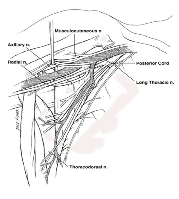
Compression of the radial nerve is most commonly described at the supinator muscle (i.e., arcade of Frohse). However, radial nerve compression can occur in the arm. Therefore, the purpose of this article is to review both etiologies of radial nerve entrapment and the sites at which this can occur in the arm. The clinical presentation of radial nerve entrapment in the arm and how it differs from that of entrapment at other sites is reviewed and the conditions potentially predisposing to nerve entrapment are described. Particular attention is paid to the nerve's course and potential variants of the anatomical structures in the arm. In each case, the recommended course of management for the neuropathy is described. Injury of the radial nerve can arise from a varied set of pathologies including trauma, tumors, anomalous muscles, and intramuscular injections. Physicians should have a good working knowledge of the anatomy and potential mechanisms for radial nerve injury.

KEYWORDS:

anatomy; brachial plexus; compression; neuropathy; upper limb

Anterolateral Triangle: A Cadaveric Study with Neurosurgical Significance.

Cureus. 2018 Feb 12;10(2):e2185. doi: 10.7759/cureus.2185.

Granger ABricoune ORajnauth TKimball DKimball HTubbs RSLoukas M.


Abstract

The anterolateral triangle is one of 10 surgical triangles of the cavernous sinus and serves as an important anatomic landmark for the skull base surgeon. There are few studies in the English literature that have precisely defined and measured the borders of the anterolateral triangle and little agreement has been made regarding the nomenclature within the English literature. A total of 12 midsagittally hemisected adult human cadaveric head halves were dissected to expose the anterolateral triangle. The triangle was defined and measurements of the anterior, posterior, and lateral borders were taken. The mean lengths and standard deviations of the anterior, posterior, and lateral borders were 8.3 ± 2.2 mm, 5.9 ± 2.0 mm, and 11.5 ± 2.9 mm, respectively. The mean area and standard deviation were 20.46 ± 9.30 mm2. The anterolateral triangle is helpful in understanding and planning surgical approaches to the cavernous sinus and middle cranial fossa. As such, normal anatomic relationships and the sizes of the anterolateral triangle must first be recognized to better access the pathologic changes within and around this region.

KEYWORDS:

anterolateral triangle; dimensions; far lateral; lateralmost; neurosurgery

Imaging Characteristics of Dural Arteriovenous Fistulas Involving the Vein of Galen: A Comprehensive Review.

Cureus. 2018 Feb 11;10(2):e2180. doi: 10.7759/cureus.2180.

Kassem MWChoi PJIwanaga JMortazavi MMTubbs RS.


Abstract

Vein of Galen aneurysmal malformation (VGAM) is a rare angiopathy, which most commonly presents in infancy. Although very rare, it is associated with high morbidity and mortality rates. In order to minimize such morbid rates, a prompt diagnosis followed by a timely initiation of management is crucial. Multiple antenatal and postnatal imaging techniques for the diagnosis have been described and discussed in the literature. However, to our knowledge, a comprehensive review exploring such a list of imaging options for VGAM has never been established. We aim to review the diagnostic tools to aid in better understanding of the investigative modalities physicians may choose from when treating patients with a VGAM.

KEYWORDS:

aneurysmal malformation; arteriovenous fistula; diagnosis; imaging; prenatal; vein of galen

Cranial Nerve Foramina Part I: A Review of the Anatomy and Pathology of Cranial Nerve Foramina of the Anterior and Middle Fossa.

Cureus. 2018 Feb 8;10(2):e2172. doi: 10.7759/cureus.2172.

Edwards BWang JMIwanaga JLoukas MTubbs RS.


Abstract

Cranial nerve foramina are integral exits from the confines of the skull. Despite their significance in cranial nerve pathologies, there has been no comprehensive anatomical review of these structures. Owing to the extensive nature of this topic, Part I of our review, presented here, focuses on the foramina of the anterior and middle cranial fossae, discussing each foramen's shape, orientation, size, surrounding structures, and structures that traverse them. Furthermore, by comparing the size of each foramen against the cross-sectional areas of its contents, we estimate the amount of free space in each. We also review lesions that can obstruct the foramina and discuss their clinical consequences.

KEYWORDS:

anterior fossa; cranial nerve; cribiform plate; foramen ovale; foramen rotundum; foramina; middle fossa; optic canal; sphenoid; superior orbital fissure

Constriction of the Stomach by an Unusual Peritoneal Band.

Cureus. 2018 Feb 3;10(2):e2148. doi: 10.7759/cureus.2148.

Kassem MWPatel MIwanaga JLoukas MTubbs RS.


Abstract

Compression of intraabdominal contents can occur due to anomalous congenital bands. Herein, we describe, to our knowledge, the first case of compression of the stomach by an anomalous band extending from the lesser omentum to the greater omentum. Relevant literature is reviewed and the clinical implications of such a case are described.

KEYWORDS:

ladd's bands; omentum; peritoneal bands; stomach constriction

Some of the earliest depictions of the human spine: a glimpse into European history.

Childs Nerv Syst. 2018 Feb;34(2):179-181. doi: 10.1007/s00381-017-3419-9. Epub 2017 Apr 25.

Oakes PCKirkpatrick CChapman JROskouian RJTubbs RS


Abstract

INTRODUCTION:

Drawings of the human form have a history almost as old as mankind itself. However, illustrations of the human spine as seen with the vertebral column were not seen until much later. This paper reviews some of the early European depictions of the human vertebral column from the twelfth (e.g., Fünfbilderserie "Bone-Man": 1152 A.D.) and thirteenth (e.g., Ashmole 1292) centuries. Man's understanding of his body has evolved over hundreds of years.

CONCLUSIONS:

This glimpse into our past and early drawings of the human spine illustrate how this particular anatomical structure was perceived almost a millennium ago and would not be structurally correct renditions until Leonardo da Vinci in the fifteenth century.

KEYWORDS:

Anatomy; Art; Drawings; European; History; Vertebral column

Landmarks for Identifying the Suprascapular Foramen Anteriorly: Application to Anterior Neurotization and Decompressive Procedures.

Oper Neurosurg (Hagerstown). 2018 Feb 1;14(2):166-170. doi: 10.1093/ons/opx096.

Manouvakhova OVMacchi VFries FNLoukas MDe Caro ROskouian RJSpinner RJTubbs RS.


Abstract

BACKGROUND:

Additional landmarks for identifying the suprascapular nerve at its entrance into the suprascapular foramen from an anterior approach would be useful to the surgeon.

OBJECTIVE:

To identify landmarks for the identification of this hidden site within an anterior approach.

METHODS:

In 8 adult cadavers (16 sides), lines were used to connect the superior angle of the scapula, the acromion, and the coracoid process tip thus creating an anatomic triangle. The suprascapular nerve's entrance into the suprascapular foramen was documented regarding its position within this anatomical triangle. Depths from the skin surface and specifically from the medial-most point of the clavicular attachment of the trapezius to the suprascapular nerve's entrance into the suprascapular foramen were measured using calipers and a ruler. The clavicle was then fractured and retracted superiorly to verify the position of the nerve's entrance into the suprascapular foramen.

RESULTS:

From the trapezius, the nerve's entrance into the foramen was 3 to 4.2 cm deep (mean, 3.5 cm). The mean distance from the tip of the corocoid process to the suprascapular foramen was 3.8 cm. The angle best used to approach the suprascapular foramen from the surface was 15° to 20°.

CONCLUSION:

Based on our study, an anterior suprascapular approach to the suprascapular nerve as it enters the suprascapular foramen can identify the most medial fibers of the trapezius attachment onto the clavicle and insert a finger at an angle of 15° to 20° laterally and advanced to an average depth of 3.5 cm.

Copyright © 2017 by the Congress of Neurological Surgeons

KEYWORDS:

Brachial plexus; Nerve entrapment; Nerve repair; Neurotization; Shoulder

Potential Mechanism for Some Postoperative C5 Palsies: An Anatomical Study.

Spine (Phila Pa 1976). 2018 Feb 1;43(3):161-166. doi: 10.1097/BRS.0000000000002281.

Alonso FVoin VIwanaga JHanscom DChapman JROskouian RJLoukas MTubbs RS.


Abstract

STUDY DESIGN:

Anatomical Study.

OBJECTIVE:

Determine if shoulder depression (eg, taping the shoulders) might result in C5 nerve traction and subsequent injury.

SUMMARY OF BACKGROUND DATA:

Postoperative C5 nerve palsy is a recognized entity that is still often enigmatic. Inferior shoulder depression is usually employed to assist with surgical visualization during cervical spine procedures.

METHODS:

In the supine position, 10 adult fresh frozen human cadavers underwent dissection of the spinal cord and its adjacent dorsal, ventral roots, and spinal nerves from C4 to T1. In the supine position, the head was rotated ipsilaterally, contralaterally, and in lateral flexion. The shoulder was elevated, retracted, protracted, and depressed all with direct observation of nerve roots, intradural ventral/dorsal rootlets, or the spinal cord. The effects of these movements upon the cervical nerve rootlets were measured.

RESULTS:

The greatest displacement of nervous tissue was generated by shoulder depression and occurred primarily at the intradural rootlet level. The nerve rootlets that underwent the greatest average displacement were found at C5, with a decreasing gradient to C7 and no gross motion at C8 or T1. With maximal shoulder depression, C5-C7 rootlet tension produced cord movement to the ipsilateral side, touching the dura mater covering the lateral vertebral column with the C5 nerve root moving farthest.

CONCLUSION:

Shoulder depression is often used during cervical spine surgery. In cadavers, shoulder depression causes significant tension and displacement of the C5 nerve rootlets, and in the extreme, cord displacement to the ipsilateral side. This could be a mechanism for injury, putting patients at greater risk for postoperative C5 palsy.

LEVEL OF EVIDENCE:

5.

Guest UserComment
Anatomical study of the palatine aponeurosis: application to posterior palatal seal of the complete maxillary denture.

Surg Radiol Anat. 2018 Feb;40(2):179-183. doi: 10.1007/s00276-017-1911-2. Epub 2017 Aug 19.

Iwanaga JKido JLipski MTomaszewska IMTomaszewski KAWalocha JAOskouian RJTubbs RS.


Abstract

The palatine aponeurosis is a thin, fibrous lamella comprising the extended tendons of the tensor veli palatini muscles, attached to the posterior border and inferior surface of the palatine bone. In dentistry, the relationship between the "vibrating line" and the border of the hard and soft palate has long been discussed. However, to our knowledge, there has been no discussion of the relationship between the palatine aponeurosis and the vibrating line(s). Twenty sides from ten fresh frozen White cadaveric heads (seven males and three females) whose mean age at death was 79 years) were used in this study. The thickness of the mucosa including the submucosal tissue was measured. The maximum length of the palatine aponeurosis on each side and the distance from the posterior nasal spine to the posterior border of the palatine aponeurosis in the midline were also measured. The relationship between the marked borderlines and the posterior border of the palatine bone was observed. The thickness of the mucosa and submucosal tissue on the posterior nasal spine and the maximum length of the palatine aponeurosis were 3.4 mm, and 12.2 mm on right side and 12.8 mm on left, respectively. The length of the palatine aponeurosis in the midline was 4.9 mm. In all specimens, the borderline between the compressible and incompressible parts corresponded to the posterior border of the palatine bone.

KEYWORDS:

Anatomy; Aponeurosis; Cadaver; Complete denture; Soft palate

The Alar Ligaments: A Cadaveric and Radiologic Comparison Study.

World Neurosurg. 2018 Feb;110:517-520. doi: 10.1016/j.wneu.2017.07.080.

Sardi JPIwanaga JVoin VSchmidt CLoukas MChapman JROskouian RJTubbs RS.


Abstract

OBJECTIVE:

A precise anatomical description of the alar ligaments is important to better understand their biomechanical and pathologic implications. Although there are several studies regarding their anatomy, the literature is inconsistent. To our knowledge, there are no reports that compare cadaveric morphologic findings with computed tomography (CT) images of the alar ligaments.

METHODS:

Eight sides from 4 fresh-frozen cadaveric specimens were used in this study. After routine dissection of the craniocervical junction, the alar ligaments were exposed. We carried out measurements of the alar ligaments, their position within the craniovertebral junction, and their relation to the dens and adjacent structures. Fine-cut CT of the specimens was performed, and the measurements were later compared with the original cadaveric dissections.

RESULTS:

Alar ligaments were attached to the upper half of the lateral surface of the dens and ran laterally to its insertion just medial to the occipital condyle. The ligaments were found to have an ovoid cross-sectional area with a nearly horizontal caudocranial trajectory and comparable diameters in both anteroposterior and superoinferior directions between the CT and cadaveric measurements.

CONCLUSIONS:

There were small but not statistically significant differences in the measurements between the cadaver specimens and the CT images. There was however, a strong correlation between the proximal and distal insertions, as well as the orientation of the fibers, that suggests CT images can be an appropriate approach to the study of the anatomical and 3-dimensional features of the alar ligaments.

Copyright © 2017 Elsevier Inc. All rights reserved.

KEYWORDS:

Alar ligaments; Anatomy; Occipito-atlantoaxial complex; Spine; Tomography

Cervical Rib Prevalence and its Association with Thoracic Outlet Syndrome: A Meta-Analysis of 141 Studies with Surgical Considerations.

World Neurosurg. 2018 Feb;110:e965-e978. doi: 10.1016/j.wneu.2017.11.148. Epub 2017 Dec 2.

Henry BMVikse JSanna BTaterra DGomulska MPękala PATubbs RSTomaszewski KA.

Abstract

BACKGROUND:

Cervical ribs (CR) are supernumerary ribs that arise from the seventh cervical vertebra. In the presence of CR, the boundaries of the interscalene triangle can be further constricted and result in neurovascular compression and thoracic outlet syndrome (TOS). The aim of our study was to provide a comprehensive evidence-based assessment of CR prevalence and their association with TOS as well as surgical approach to excision of CR and surgical patients' characteristics.

METHODS:

A thorough search of major electronic databases was conducted to identify any relevant studies. Data on the prevalence, laterality, and side of CR were extracted from the eligible studies for both healthy individuals and patients with TOS. Data on the type of TOS and surgical approach to excision of CR were extracted as well.

RESULTS:

A total of 141 studies (n = 77,924 participants) were included into the meta-analysis. CR was significantly more prevalent in patients with TOS than in healthy individuals, with pooled prevalence estimates of 29.5% and 1.1%, respectively. More than half of the patients had unilateral CR in both the healthy and the TOS group. The analysis showed that 51.3% of the symptomatic patients with CR had vascular TOS, and 48.7% had neurogenic TOS. Most CR were surgically excised in women using a supraclavicular approach.

CONCLUSIONS:

CR ribs are frequent findings in patients with TOS. We recommended counseling asymptomatic patients with incidentally discovered CR on the symptoms of TOS, so that if symptoms develop, the patients can undergo prompt and appropriate workup and treatment.

Copyright © 2017 Elsevier Inc. All rights reserved.

KEYWORDS:

Anatomy; Cervical rib; Meta-analysis; Thoracic outlet syndrome

Rare Concurrent Retroclival and Pan-Spinal Subdural Empyema: Review of Literature with an Uncommon Illustrative Case.

World Neurosurg. 2018 Feb;110:326-335. doi: 10.1016/j.wneu.2017.11.082. Epub 2017 Nov 23.

Mortazavi MMQuadri SASuriya SSFard SAHadidchi SAdl FHArmstrong IGoldman RTubbs RS.


Abstract

BACKGROUND:

Subdural empyema can present as a spinal subdural empyema (SSE) or a cranial subdural empyema (CSE). Although they differ somewhat in epidemiology, etiology, pathophysiology, and symptomatology and occur separately, they rarely manifest together. The aim of this article is to review the literature concerning the clinical presentation, clinical course, and treatment options for managing concurrently occurring SSE and CSE.

METHODS:

The literature in the Medline database was reviewed with key words including but not limited to subdural empyema, retroclival empyema, and Streptococcus mitis. No similar reports were found in the database involving infection with this type of microorganism in this anatomical region.

RESULTS:

Only 3 cases with concurrent CSE and SSE were found in the literature caused by various etiologic agents. Two of the patients recovered with no neurologic deficit, whereas one fatality was reported. One new illustrative case caused by Streptococcus mitis is also presented.

CONCLUSIONS:

CSE and SSE are neurosurgical emergencies, often requiring prompt surgical evacuation. Although very rare, Streptococcus mitis can cause spinal subdural empyema or retroclival abscesses. Natural history of this disease is grave without treatment. Delays in diagnosis and treatment are directly related to mortality and severe morbidity in patients with intracranial and spinal subdural empyema. Prompt recognition and treatment are essential to preclude severe neurologic disabilities or in rare cases a fatal outcome. A treatment paradigm for cranio-spinal empyema is proposed.

Copyright © 2017 Elsevier Inc. All rights reserved.

KEYWORDS:

Cranial subdural empyema; Retroclival empyema; Spinal subdural empyema; Streptococcus mitis; Subdural empyema

Rapid Progression of Ossification of the Posterior Longitudinal Ligament After Anterior Cervical Discectomy and Fusion.

World Neurosurg. 2018 Feb;110:11-16. doi: 10.1016/j.wneu.2017.10.105. Epub 2017 Oct 28.

Rustagi TAlonso FSchmidt COskouian RJChapman JRTubbs RSFisahn C.


Abstract

BACKGROUND:

Ossification of the posterior longitudinal ligament (OPLL) has a reported incidence of 1.9%-4.3%. Disease progression is associated with surgery, with most studies focusing on OPLL progression after laminoplasty. The continued range of motion following surgery is believed to place strain on adjacent levels, driving calcification of the ligament. We present a case of marked progression of OPLL at levels adjacent to a previous anterior cervical discectomy and fusion.

CASE REPORT:

A 59-year-old man initially presented for progressive loss of balance and dexterity and underwent a C4-6 anterior cervical discectomy and fusion procedure. Computed tomography performed 1 year postoperatively showed fusion across C4-6 with no evidence of OPLL at any level. Two years following index surgery, the patient reported right-side arm pain. Computed tomography revealed new minimal OPLL opposite the C3-4 level that was not causing cord compression. A sparing midline bilateral C3-7 laminotomy was performed to correct stenosis seen on magnetic resonance imaging. At 7 years after the index procedure, the patient presented with myelopathic symptoms exceeding symptoms at his index presentation. Computed tomography revealed marked progression of OPLL. The patient underwent C2-T2 posterior fusion with laminectomy from C2-C7 and responded well with gradual improvements in balance and arm pain after surgery.

CONCLUSIONS:

Our report demonstrates that OPLL progression, which is largely reported following laminoplasty, may similarly occur following anterior cervical discectomy and fusion and supports the concept of motion-related OPLL progression.

Copyright © 2017 Elsevier Inc. All rights reserved.

KEYWORDS:

ACDF; Cervical fusion; Laminoplasty; Myelopathy; OPLL

Anatomic Study of Extracranial Needle Trajectory Using Hartel Technique for Percutaneous Treatment of Trigeminal Neuralgia.

World Neurosurg. 2018 Feb;110:e245-e248. doi: 10.1016/j.wneu.2017.10.140. Epub 2017 Nov 22.

Iwanaga JBadaloni FLaws TOskouian RJTubbs RS.


Abstract

OBJECTIVE:

The aim of this study was to describe the anatomic trajectory of the extracranial needle for percutaneous rhizotomy and correlate this with structures at risk during such a procedure.

METHODS:

Six sides from 3 frozen fresh Caucasian heads were used in this study. Hartel anatomic landmarks for percutaneous trigeminal rhizotomy procedures were used. A free hand technique was then used, and intraprocedural visualization of the needle was performed with fluoroscopy. When the procedure was completed, the face was dissected along with the needle pathway up to the foramen ovale in order to evaluate for any damaged structures.

RESULTS:

On all sides, the needle passed lateral to the buccinator muscle and near the parotid duct to then pierce the superior head of the lateral pterygoid muscle to enter the infratemporal fossa. This placed the needle near the buccal nerve on all sides, although no direct injury was noted. Although very near, no branches of the facial nerve or artery were damaged. On 1 side, the maxillary artery was pierced.

CONCLUSIONS:

To our knowledge, this is the first study to describe the detailed extracranial anatomic needle pathway using the Hartel approach. Such data might help surgeons better recognize potential complications from such procedures.

Copyright © 2017 Elsevier Inc. All rights reserved.

KEYWORDS:

Anatomy; Cadaver; Foramen ovale; Neurosurgery; Trigeminal neuralgia

Spontaneous Fusion of S2/S3 Spondyloptosis in an Adult.

World Neurosurg. 2018 Feb;110:129-132. doi: 10.1016/j.wneu.2017.10.001. Epub 2017 Oct 12.

Vahedi PRymarczuk GNGillick JLTubbs RSWilson JPrasad SK.


Abstract

BACKGROUND:

Spondyloptosis is grade V on the Meyerding classification. Traumatic spondyloptosis can occur throughout the spinal column, particularly at junctional levels, and finding an ideal surgical strategy to address it remains a challenge for spinal surgeons. The sacrum is considered a united bone in adults, and sacral intersegmental spondyloptosis is extremely rare.

CASE REPORT:

Herein, we present an unusual case of S2/S3 spondyloptosis in a 27-year-old female patient with spontaneous solid fusion.

CONCLUSIONS:

This case demonstrates that similar distal sacral pathologies may be managed conservatively when there is no associated neurologic deficit, and the osteodiskoligamentous integrity of the lumbosacropelvic unit remains intact. Our report plus the very few published papers in the literature illustrate the natural history of uncomplicated traumatic spondyloptosis and support the role of in situ fusion and instrumentation as a reliable alternative to circumferential fusion in patients who cannot tolerate staged or prolonged operations.

Copyright © 2017. Published by Elsevier Inc.

KEYWORDS:

Fusion; Sacral; Spondyloptosis

Complications of Subcutaneous Contraception: A Review.

Cureus. 2018 Jan 31;10(1):e2132. doi: 10.7759/cureus.2132.

Ramdhan RCSimonds EWilson CLoukas MOskouian RJTubbs RS.


Abstract

Over 62 million women in the United States are of childbearing age and 60% of them use contraception. Subcutaneous contraceptives include implantable contraceptives and subcutaneous injections. Implantable contraception involves subdermal time-release of synthetic progestin, which allows for several years of continuous, highly effective contraception. Its main effects are inhibition of ovulation and thickening of the cervical mucus. Many complications have been associated with subcutaneous contraception, including menstrual disturbances, headache, weight gain, acne, dizziness, mood disturbances, nausea, lower abdominal pain, hair loss, loss of libido, pain at the implant site, neuropathy, and follicular cysts. Using standard search engines, the complications of subcutaneous contraception are reviewed. Patients should be adequately counseled on the possible complications and side effects of subcutaneous contraception to help them make an informed decision when choosing the right contraceptive to meet their needs.

KEYWORDS:

contraceptives; neuropathy; subcutaneous; subdermal

Anatomical Study of the Extreme Lateral Transpsoas Lumbar Interbody Fusion with Application to Minimizing Injury to the Kidney.

Cureus. 2018 Jan 29;10(1):e2123. doi: 10.7759/cureus.2123.

Iwanaga JYilmaz ETawfik TAbdul-Jabbar AVetter MMoisi MWatanabe KYamaki KITubbs RSOskouian RJ.


Abstract

Objective Since the extreme lateral lumbar interbody fusion procedure was first reported by Ozgur in 2006, a large number of clinical studies have been published. Anatomical studies which explore methods to avoid visceral structures, such as the kidney, with this approach have not been examined in detail. We dissected the retroperitoneal space to analyze how the extreme lateral transpsoas approach to the lumbar spine could damage the kidney and related structures. Methods Eight sides from four fresh Caucasian cadavers were used for this study. The latissimus dorsi muscle and the thoracolumbar fascia were dissected to open the retroperitoneum. The fat tissue was removed. Steel wires were then put into the intervertebral disc spaces. Finally, the closest distance between kidney and wires on each interdiscal space was measured. Results The closest distance from the wire in the interdiscal space on L1/2, L2/3 and L3/4 to the kidney ranged from 13.2 mm to 32.9 mm, 20.0 mm to 27.7 mm, and 20.5 mm to 46.6 mm, respectively. The distance from the kidney to the interdiscal space at L4/5 was too great to be considered applicable to this study. Conclusions The results of this study might help surgeons better recognize the proximity of the kidney and avoid injury to it during the extreme lateral transpsoas approach to the lumbar spine.

KEYWORDS:

anatomy; complications; spine surgery; surgery; vertebral column