External cortical landmarks for localization of the hippocampus: Application for temporal lobectomy and amygdalohippocampectomy.

Surg Neurol Int. 2018 Aug 22;9:171. doi: 10.4103/sni.sni_446_17. eCollection 2018.

Tubbs RSLoukas MBarbaro NMShah KJCohen-Gadol AA.


Abstract

BACKGROUND:

Accessing the hippocampus for amygdalohippocampectomy and minimally invasive procedures, such as depth electrode placement, require an accurate knowledge regarding the location of the hippocampus.

METHODS:

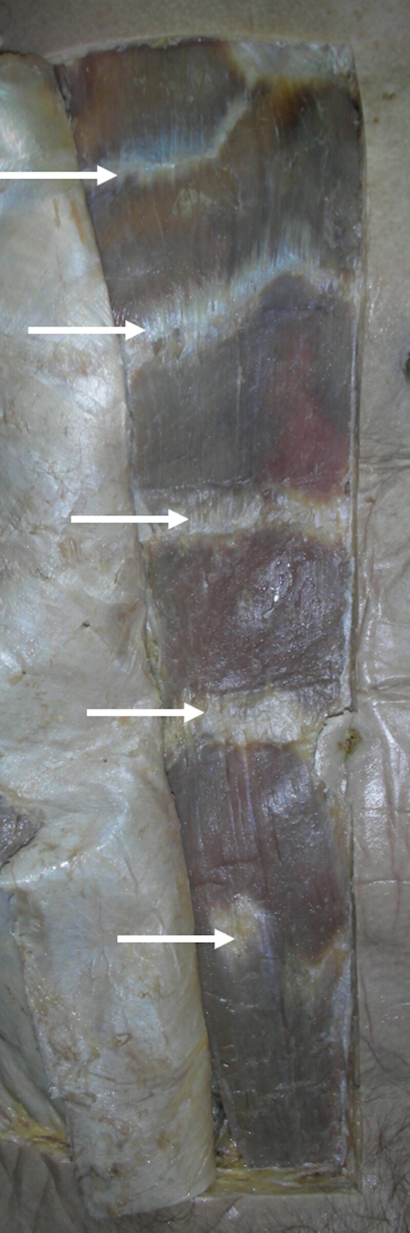
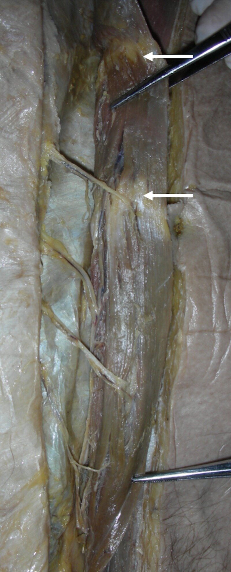
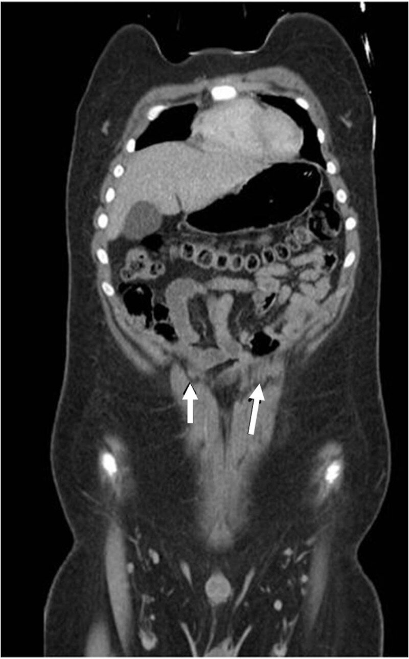
The authors removed 10 human cadaveric brains from the cranium and observed the relationships between the lateral temporal neocortex and the underlying hippocampus. They then measured the distance between the hippocampus and superficial landmarks. The authors also validated their study using magnetic resonance imaging (MRI) scans of 10 patients suffering from medial temporal lobe sclerosis where the distance from the hippocampal head to the anterior temporal tip was measured.

RESULTS:

In general, the length of the hippocampus was along the inferior temporal sulcus and inferior aspect of the middle temporal gyrus. Although the hippocampus tended to be more superiorly located in female specimens and on the left side, this did not reach statistical significance. The length of the hippocampus tended to be shorter in females, but this too failed to reach statistical significance. The mean distance from the anterior temporal tip to the hippocampal head was identical in the cadavers and MRIs of patients with medial temporal lobe sclerosis.

CONCLUSIONS:

Additional landmarks for localizing the underlying hippocampus may be helpful in temporal lobe surgery. Based on this study, there are relatively constant anatomical landmarks between the hippocampus and overlying temporal cortex. Such landmarks may be used in localizing the hippocampus during amygdalohippocampectomy and depth electrode implantation in verifying the accuracy of image-guided methods and as adjuvant methodologies when these latter technologies are not used or are unavailable.

KEYWORDS:

Anatomy; epilepsy surgery; hippocampectomy; landmarks; neurosurgery; temporal lobe

Bifid Xiphoid Process: Case Report and Review.

Cureus. 2018 Aug 16;10(8):e3153. doi: 10.7759/cureus.3153.

Eid SIwanaga JOskouian RJLoukas MTubbs RS.


Abstract

The xiphoid process is a bony process that comprises part of the sternum. This anatomical structure exhibits several morphological variations, which may complicate diagnostic examinations and invasive thoracic procedures. Variations include bifurcated or trifurcated, deflected, and curved processes. This report discusses a case of a bifid xiphoid process during cadaveric dissection and compares similar findings in the literature. The aim is to improve our knowledge of anatomical variations in an effort to reduce complications and improve patient care.

KEYWORDS:

anatomy; bifid xiphoid process; variation; xiphoid process


Intelligence Quotient in Patients with Myelomeningocele: A Review.

Cureus. 2018 Aug 13;10(8):e3137. doi: 10.7759/cureus.3137.

Alimi YIwanaga JOskouian RJLoukas MTubbs RS.


Abstract

It has been proposed that hydrocephalus in children with myelomeningocele (MMC) can indicate a low intelligence quotient (IQ). Others have argued that it is not the mere presence of hydrocephalus but the superimposition of cerebrospinal fluid (CSF) infections, multiple shunt procedures, and other CNS complications that lead to the lowering of IQ in these patients. In this paper, we review the literature to clarify the information about IQ in patients with MMC and whether it changes after infections and shunt procedures. We have also considered the other factors that could be involved in the IQ development of these patients and the differences revealed by the brain imaging of individuals with MMC. The consensus remains that patients with MMC, with or without complications, tend to have a lower IQ than those without MMC. Hydrocephalus appears to decrease the IQ further in MMC patients. Some have proposed that prenatal repair of the MMC lesion reduces the need for ventricular shunting after birth, thus decreasing the risk of shunt complications such as a CNS infection, which can have a negative effect on IQ. More studies are needed to assess other risk factors (apart from folate deficiency) and genetic factors that could contribute to the development of MMC and their possible effects on patient IQ.

KEYWORDS:

hydrocephalus; intelligence quotient; myelomeningocele; spina bifida; venticuloperitoneal shunt

Wormian Bone of the Orbit: A Case Report.

Cureus. 2018 Aug 7;10(8):e3117. doi: 10.7759/cureus.3117.

Rizvi AIwanaga JOskouian RJLoukas MTubbs RS.


Abstract

Wormian bones are formed due to abnormal ossification centers in various locations in the skull. Genetic and/or environmental factors have been proposed to explain their formation. These bones can be normal anatomical variants or associated with a number of pathological conditions. The literature reports the most common locations of these bones as the cranial sutures, and reports of the presence of these bones in the orbit are rare. Clinically, these bones in the orbit can simulate fractures on imaging or can dislodge during surgery causing injury to the surrounding structures. Herein, we report a case of wormian bones of the orbit and discuss other reports from the literature.

KEYWORDS:

cranial sutures; fontanelles; imaging; orbit; orbital fractures; supernumerary bones; sutural bones; wormian bones

Additional Attachment of the Semitendinosus and Gracilis Muscles to the Crural Fascia: A Review and Case Illustration.

Cureus. 2018 Aug 7;10(8):e3116. doi: 10.7759/cureus.3116.

Rizvi AIwanaga JOskouian RJLoukas MTubbs RS.


Abstract

The semitendinosus and gracilis muscles insert primarily onto the superior medial aspect of the tibia. These tendons can be harvested for anterior cruciate ligament reconstruction, and knowledge of their accessory attachments is important for the success of such harvesting procedures. Here, we present a case illustration and review of the attachment of these muscles into the crural fascia (deep fascia of the leg), which is often an underappreciated insertion site.

KEYWORDS:

accessory bands; acl reconstruction; crural fascia; deep fascia; gracilis; semitendinosus

Clinical Application of the IllumiScan Fluorescence Visualization Device in Detecting Oral Mucosal Lesions.

Cureus. 2018 Aug 6;10(8):e3111. doi: 10.7759/cureus.3111.

Kikuta SIwanaga JTodoroki KShinozaki KTanoue RNakamura MKusukawa J.


Abstract

OBJECTIVE:

Fluorescence visualization devices are screening devices that can be used to examine lesions of the oral mucosa non-invasively. We observed oral squamous cell carcinoma (OSCC) and leukoplakia using the IllumiScan (Shofu, Kyoto, Japan) fluorescence visualization device and examined its usefulness and characteristics.

METHODS:

We investigated 31 OSCC and nine leukoplakia in patients who were examined using the IllumiScan and treated in our department from January 2017 to February 2018. Images taken with the IllumiScan were analyzed using image analysis software. We also examined the lesions using narrowband imaging (NBI). Additionally, the IllumiScan and NBI images and the non-stained areas of iodine staining method (IOM) were visually evaluated.

RESULTS:

The average luminance of OSCC in the keratinized mucosa was significantly lower than that of OSCC in non-keratinized mucosa. The average luminance of OSCC was significantly lower than that of leukoplakia. Even in keratinized mucosa where IOM is impossible to use, the OSCC lesion exhibited fluorescence visualization loss.

CONCLUSION:

The application of the fluorescence visualization device to the oral mucosa may be useful for distinguishing between cancer and normal areas and can be used to detect OSCC in the keratinized mucosa. The use of the IllumiScan in combination with other conventional screening methods may lead to a better diagnosis.

KEYWORDS:

fluorescence visualization device; illumiscan®; leukoplakia; narrow band imaging; oral squamous cell carcinoma

Anatomical Study of the Lingual Nerve and Inferior Alveolar Nerve in the Pterygomandibular Space: Complications of the Inferior Alveolar Nerve Block.

Cureus. 2018 Aug 6;10(8):e3109. doi: 10.7759/cureus.3109.

Iwanaga JChoi PJVetter MPatel MKikuta SOskouian RJTubbs RS.


Abstract

The inferior alveolar nerve block (IANB) procedure delivers anesthetics to the pterygomandibular space through which the lingual nerve (LN) and inferior alveolar nerve (IAN) travel. Injury to the LN has been reported more often than injury to the IAN. However, the number of anatomical studies of LN injury is limited. We aimed to establish evidence by investigating LN and IAN anatomy at the level of the mandibular foramen (MF). Forty-four sides from 22 Caucasian cadaveric heads (16 fresh-frozen and six formalin-fixed cadavers) were used in this study. The LN and IAN were laterally dissected, and the diameter and the distance between the two nerves were measured at the level of the MF. The mean diameters of the LN and IAN were 2.57 mm and 2.53 mm in fresh-frozen specimens and 2.97 mm and 2.93 mm in formalin-fixed specimens, respectively. The mean diameters of the LN and IAN in all the specimens were 2.65 mm and 2.64 mm. The distance between the posterior edge of the LN and anterior edge of the IAN at the level of the MF ranged from 1.62 to 8.36 mm with a mean of 5.33 ± 1.88 mm. These findings could elucidate the risk of LN injury during the IANB procedure.

KEYWORDS:

anatomy; cadaver; dissection; inferior alveolar nerve; lingual nerve; nerve block

Tendinous Inscriptions of the Rectus Abdominis: A Comprehensive Review.

Cureus. 2018 Aug 4;10(8):e3100. doi: 10.7759/cureus.3100.

Rai R1, Azih LCIwanaga JLoukas MMortazavi MOskouian RJTubbs RS.


Abstract

The rectus abdominis muscles are interrupted by tendinous inscriptions, which typically appear as fibrous bands crossing the muscle. The current literature on tendinous inscriptions is scarce; hence, this review will provide a detailed overview of their anatomical description, including their variation, embryology, comparative anatomy, and clinical application. Understanding the anatomy and function of the tendinous inscription assists in providing clinical relevance and in guiding reconstructive surgeons in their surgical planning and design.

KEYWORDS:

anatomy; anterior abdominal wall; rectus abdominis muscle; segmentation; tendon

A Cadaveric Case of a Circular Torcular Herophili.

Cureus. 2018 Aug 4;10(8):e3099. doi: 10.7759/cureus.3099.

Lake SAltafulla JJIwanaga JOskouian RJLoukas MTubbs RS.


Abstract

The torcular Herophili is formed by the joining of the straight sinus, superior sagittal sinus, and transverse sinus. The anatomic configuration of the torcular Herophili is highly variable. In the current literature, classification systems define up to nine subtypes of the torcular Herophili. The frequency of prevalence of these anatomical variants is also variable. Herein is a case report of a circularly-shaped torcular Herophili found during cadaveric dissection.

KEYWORDS:

confluence sinus; torcular herophili; torcular morphology; torcular variants; waiting

A comprehensive review of the clivus: anatomy, embryology, variants, pathology, and surgical approaches.

Childs Nerv Syst. 2018 Aug;34(8):1451-1458. doi: 10.1007/s00381-018-3875-x. Epub 2018 Jun 28.

Rai RIwanaga JShokouhi GLoukas MMortazavi MMOskouian RJTubbs RS.


Abstract

INTRODUCTION:

The clivus is a bony structure formed by the fusion of the basioccipital and basispheniod bone at the sphenooccipital synchondrosis. This downward sloping structure from the dorsum sellae to the foramen magnum is derived from mesoderm and ectoderm properties.

METHODS:

This comprehensive review of the clivus will discuss its basic anatomy, embryology, pathological findings, and surgical implications. The clivus is an endochondral bone, formed under two processes; first, a cartilaginous base is developed, and it is secondly reabsorbed and replaced with bone. Knowledge of its embryological structure and growth of development will clarify the pathogenesis of anatomical variants and pathological findings of the clivus.

CONCLUSIONS:

Understanding the anatomy including proximity to anatomical structures, adjacent neurovasculature properties, and anatomical variants will aid neurosurgeons in their surgical management when treating pathological findings around the clivus.

KEYWORDS:

Anatomy; Clivus; Embryology; Notochord; Skull base; Surgery

Is There an Optimal Wound Closure Technique for Major Posterior Spine Surgery? A Systematic Review.

Global Spine J. 2018 Aug;8(5):535-544. doi: 10.1177/2192568218774323. Epub 2018 May 23.

Yilmaz EBlecher RMoisi MAnkush CO'Lynnger TMAbdul-Jabbar ADettori JROskouian RJ.


Abstract

STUDY DESIGN:

Systematic review.

OBJECTIVES:

In patients undergoing posterior spinal fusion: (1) What are the types and risks of wound complications in major (≥3 levels) surgery, and does the risk vary by number of levels fused? (2) What types of fascial closure result in the fewest wound complications? (3) What subcutaneous closure technique is more effective in preventing wound complications for obese patients (body mass index >30 kg/m2)? (4) What type of skin closure results in the fewest wound complications? (5) What type of dressing results in the fewest wound complications?

METHODS:

Electronic databases and reference lists of key articles were searched from January 1, 2000 to December 4, 2017 to identify studies meeting inclusion criteria.

RESULTS:

Six lower quality retrospective studies (evidence level III) met the inclusion criteria. The risk of wound complications in patients with ≥3 level posterior spine fusion ranges from 1.5% to 3.7% depending on the definition of wound complications. Skin closure with sutures resulted in fewer wound infections compared with staples (0.0% vs 8.0%, P = .023). We were unable to demonstrate an association between the number of levels fused and infection risk. Wound infections, primarily superficial, occurred less frequently with Silverlon dressing versus routine dressing.

CONCLUSIONS:

We were unable to determine if infection risk changed with increasing number of levels fused. There is a lack of evidence for optimal wound closure technique in posterior spine surgery. Several questions still remain unanswered, such as the optimal fascial closure technique or the optimal subcutaneous closure technique in obese patients.

KEYWORDS:

skin closure; spine surgery; wound closure; wound complication; wound infection

Presence of a foramen arcuale as a possible cause for headaches and migraine: Systematic review and meta-analysis.

J Clin Neurosci. 2018 Aug;54:113-118. doi: 10.1016/j.jocn.2018.05.008. Epub 2018 May 23.

Pękala PAHenry BM1, Phan KPękala JRTaterra DWalocha JATubbs RSTomaszewski KA.


Abstract

The foramen arcuale (FA) is an osseous bridge that can be formed on the posterior arch of the atlas, often observed on lateral radiographs of the cervical spine. Currently, there is a lack of studies that systematically review the available literature on the FA and its role in headaches and migraines. Therefore, we conducted an extensive search of the major electronic databases to identify all studies that reported relevant data on patients with versus without FA and prevalence of headaches or migraines. No date or language restrictions were applied. Data on the prevalence, type (complete and incomplete), and laterality of the FA and relationship with headaches or migraines were extracted and pooled into a meta-analysis. There were 168/412 (40.8%) patients with headaches in the FA group compared to 368/1691 (21.8%) patients with headaches in the no FA group (OR 4.68; P = 0.002). The proportion of headaches for complete FA was 73/125 (58.4%) compared with 80/413 (19.4%) for incomplete FA (OR 5.04; P = 0.04). When comparing incomplete FA to no FA, the proportion of headaches was 36/85 (42.3%) vs 65/560 (11.6%), and this was significantly different (OR 6.13; P = 0.009). The proportion of headaches in patients with bilateral FA was 67/132 (50.8%) compared to 80/140 (57.1%) in the unilateral group (OR 1.36; P = 0.72). We performed the first meta-analysis to demonstrate the significant association between FA and headaches, and provided further support for the notion of the clinical relevance of this anatomical variant.

Copyright © 2018 Elsevier Ltd. All rights reserved.

KEYWORDS:

Foramen arcuale; Headache; Ponticulus posticus; Vertigo

Anatomic Study of Superior Cluneal Nerves: Application to Low Back Pain and Surgical Approaches to Lumbar Vertebrae.

World Neurosurg. 2018 Aug;116:e766-e768. doi: 10.1016/j.wneu.2018.05.087. Epub 2018 May 19.

Iwanaga JSimonds EPatel MOskouian RJTubbs RS.


Abstract

OBJECTIVE:

The aim of this study was to investigate the anatomy of the superior cluneal nerves more proximal to the posterior layer of the thoracolumbar fascia.

METHODS:

Twelve sides of 6 fresh-frozen cadavers were used. The age at death ranged from 54 to 88 years. After a transverse skin incision 10 mm above the iliac crest, the superior cluneal nerves were detected by blunt dissection and traced back to the dorsal root ganglia. The diameter of the nerves from L1 to L3 was measured. Also, the relationship to the erector spinae muscle and dorsal ramus was recorded.

RESULTS:

The mean diameters of the origin of the L1, L2, and L3 were 1.71 ± 0.29 mm, 1.73 ± 0.40 mm, and 1.52 ± 0.55 mm, respectively. On 7 sides (58.3%) for L1, seven sides (58.3%) for L2, and 10 sides (83.3%) for L3, the nerves pierced the iliocostalis muscle. One side (8.3%) for L2 and one (8.3%) for L3 had no cutaneous branch.

CONCLUSIONS:

The results of this study could help to elucidate the anatomy of the superior cluneal nerves and help avoid complications during surgical approaches to the lumbar spine.

Copyright © 2018 Elsevier Inc. All rights reserved.

KEYWORDS:

Anatomy; Iliac crest; Low back pain; Lumbar vertebrae; Nerve block

Incidentally Found Double Aortic Arch While Treating Posterior Inferior Cerebellar Artery Aneurysm: Conversion from Femoral to Radial Artery Access.

World Neurosurg. 2018 Aug;116:1-4. doi: 10.1016/j.wneu.2018.04.142. Epub 2018 Apr 28.

Patel MIwananga JBentley JWang BHPatel ATubbs RSMonteith S.


Abstract

BACKGROUND:

Asymptomatic double aortic arches are a unique occurrence.

CASE DESCRIPTION:

An incidental finding of a double aortic arch in an elderly male was discovered during workup of a transient ischemic attack. The following case presentation details an effective treatment approach in cerebrovascular stenting in a patient with variant aortic arch anatomy. The initial diagnostic cerebral angiogram was performed via transfemoral approach and was quite challenging.

CONCLUSIONS:

Faced with challenging anatomy, the radial artery approach is a viable option when navigating into the cerebrovascular anatomy for stenting when proximal variants such as a double aortic arch are identified.

Copyright © 2018 Elsevier Inc. All rights reserved.

KEYWORDS:

Cerebrovascular stenting; Double aortic arch; PICA; Radial artery

Neurovascular Relationships of S2AI Screw Placement: Anatomic Study.

World Neurosurg. 2018 Aug;116:e108-e112. doi: 10.1016/j.wneu.2018.04.095. Epub 2018 Apr 22.

Abdul-Jabbar AYilmaz EIwanaga JTawfik TO'Lynnger TMSchildhauer TAChapman JOskouian RJTubbs RS.


Abstract

INTRODUCTION:

The S2 alar-iliac (S2AI) screw is a modification of the traditional iliac fixation technique and has surgical and biomechanical benefits. However, there are significant regional neurovascular structures along the path of such screws. Therefore the current anatomic study was performed to better elucidate these relationships.

METHODS:

Using fluoroscopy, S2AI screws were placed in 2 adult cadavers through a standard posterior midline exposure. The screw insertion point was placed 10 mm lateral to a line bisecting the S1 and S2 foramina, adjacent to the sacroiliac joint. Using 30- to 40-degree lateral angulation from the midline and 20- to 30-degree caudal angulation, a pedicle probe was directed toward the anterior inferior iliac spine. The final trajectory was positioned to sit 1-2 cm superior to the greater sciatic foramen. Lastly, the screws and surrounding bone were drilled in order to visualize both lateral and medial neurovascular relationships.

RESULTS:

Removing the bone around the S2AI-screw illustrated the close relationship to the medial (internal) neurovascular structures including the obturator nerve, lumbosacral trunk, sacral plexus and, specifically, the S1 ventral ramus and iliac vein and artery. By removing the outer cortex of the ilium, the close relationship to the superior gluteal artery, vein, and nerve was observed. In addition, we were able to identify the proximity to the iliopsoas muscle and internal iliac vessels.

CONCLUSIONS:

A comprehensive knowledge of the surrounding neurovascular anatomy relevant to S2AI screw placement can decrease patient morbidity and allow spine surgeons to better diagnose potential postoperative complications.

Copyright © 2018 Elsevier Inc. All rights reserved.

KEYWORDS:

Anatomy; Complications; Pelvis; S2AI; Spine; Spinopelvic fixation; Surgery

Review of Treatment of Gunshot Wounds to Head in Late 19th Century.

World Neurosurg. 2018 Aug;116:396-401. doi: 10.1016/j.wneu.2018.04.043. Epub 2018 Apr 17.

Protas MYilmaz EPatel APIwanaga JOskouian RJTubbs RS.


Abstract

INTRODUCTION:

During the late 19th century, the seeds of modern neurosurgery were planted to bloom into what it is now known. Wars such as the American Civil War and Crimean War drove the need to find better ways of preventing mortality from gunshot wounds to the head. However, the mortality rate from all major surgical procedures to the head, neck, and face remained staggering. Herein, we describe the surgical treatments for head and neck injuries in order to improve our understanding of neurosurgical procedures performed during the late 19th century.

METHODS:

A literature search was conducted using PubMed and Google Books for available articles pertaining to treatment for gunshot wounds to the head during the 19th century. Search terms included "Gunshot wounds, Treatment, Civil War," "Gunshot wound, Treatment 19th century," and "Gunshot wounds, Treatment, 1800s." Literature was excluded if not in English or if no translation was provided. Most of the information was taken from the International Encyclopedia of Surgery Volume II.

RESULTS:

Surgical care for gunshot wounds to the cranium were based on depth and involved finding the bullet, controlling the bleeding, and preventing further brain injury. Surgical treatment for a gunshot wound to the face or neck involved controlling the bleeding, with a focus on maintaining the airway.

CONCLUSIONS:

Because of improved understanding of infectious processes and technologic advances in surgical equipment, the late 19th century was a major milestone in creating modern day neurosurgery. The methodology behind today's treatments is no different from that of the late 19th century.

Copyright © 2018 Elsevier Inc. All rights reserved.

KEYWORDS:

Civil war; Gunshot wounds; Head injury; Surgery

The Tentorium Cerebelli: A Comprehensive Review Including Its Anatomy, Embryology, and Surgical Techniques.

Cureus. 2018 Jul 31;10(7):e3079. doi: 10.7759/cureus.3079.

Rai RIwanaga JShokouhi GOskouian RJTubbs RS.


Abstract

The tentorium cerebelli functions as a partition, dispelling the burden of weight from supratentorial structures upon inferior brain matter. Clinicians and neurosurgeons, when assessing pathological findings, should have knowledge regarding the tentorium cerebelli anatomy. This work of literature is a comprehensive review of the tentorium cerebelli, including its anatomy, embryology, and clinical and surgical implications. The evolutionary pattern demonstrates sequential stages to higher mammalian lineage. An understanding of the complexity of the neurovascular structures and the anatomy of the tentorium cerebelli is crucial for surgical procedures by neurosurgeons.

KEYWORDS:

dural sinus; embryology; incisura; tentorial notch; tentorium cerebelli

The Incisive Canal: A Comprehensive Review.

Cureus. 2018 Jul 30;10(7):e3069. doi: 10.7759/cureus.3069.

Lake SIwanaga JKikuta SOskouian RJLoukas MTubbs RS.


Abstract

The incisive canal, also known as the nasopalatine canal, is an interosseous conduit through the anterior maxilla connecting the oral and nasal cavities. Within this canal lies the nasopalatine nerve and the vascular anastomosis between the greater palatine and sphenopalatine arteries. The embryology of the canal has led to interesting theories explaining its function. Efforts have been made to describe the morphometrics of the incisive canal by radiologic evaluation across sex and ethnicities. This paper aims to review the current literature on the embryology, anatomy, and clinical relevance of the incisive canal.

KEYWORDS:

dental implant; incisive canal; incisive canal cysts; maxillary incisive canal; nasopalatine canal; nasopalatine nerve block

The Course of the V2 Segment of the Vertebral Arteries in Klippel-Feil Syndrome: A Case Report.

Cureus. 2018 Jul 24;10(7):e3038. doi: 10.7759/cureus.3038.

Rizvi AIwanaga JOskouian RJLoukas MTubbs RS.


Abstract

Klippel-Feil syndrome is a congenital disorder characterized by the fusion of one or more cervical vertebrae leading to limitations in the rotation, extension, and flexion of the neck and possible neurological symptoms. Other abnormalities can also be found in these patients. The anatomy of the vasculature can be abnormal in these patients including variations in the course and origin of the vertebral arteries potentially leading to intra-operative complications. Herein, we report a case of Klippel-Feil syndrome and detail the course of the vertebral arteries in an osteological specimen.

KEYWORDS:

cervical vertebrae; ct angiography; imaging; klippel-feil syndrome; surgical management; vertebral artery