Radial Artery Injury with Attempted Intravenous Catheterization on the Dorsal Hand: A Case Report.

Cureus. 2018 Jul 23;10(7):e3033. doi: 10.7759/cureus.3033.

Alimi YIwanaga JOskouian RJLoukas MTubbs RS.


Abstract

Intravenous (IV) access on the dorsum of the hand is of high clinical significance. As it is possible for an arterial injury during IV access on the dorsum of the hand to occur, clinicians and healthcare providers should be cognizant of regional anatomy and anatomical variations during IV placement so as to prevent injuries to the radial artery. We report a case of an injury to the radial artery with an attempted hand IV placement in a cadaver and suggest possible ways to prevent this complication.

KEYWORDS:

anatomy; injury; intravenous; radial artery

Brain Herniation Through the Cribriform Plate: Review and Comparison to Encephaloceles in the Same Region.

Cureus. 2018 Jul 10;10(7):e2961. doi: 10.7759/cureus.2961.

Rai RIwanaga JLoukas MOskouian RJTubbs RS.


Abstract

Herniations of the brain and/or meninges through an opening of the skull often occur through the foramen magnum, e.g., Chiari malformations and encephaloceles. The herniation of brain matter through the cribriform plate is a rare incident and has not been reported frequently. The presence of such an occurrence still requires attention and anatomical understanding. This review will examine the potential causes of cribriform plate herniation and its distinguishability to nasal encephaloceles. The sloping of brain tissue toward potential space/opening in response to elevated pressures in the cranium to accommodate for the added pressure are features seen in herniation. The presence of a pedicle and stalk seen in an encephalocele define its characteristics, which are not visible in a 'classical' herniation. Cerebrospinal fluid (CSF) fistula commonly occurs at the cribriform plate, and due to the structural weakness, a pathway is formed. This is often seen in conjunction with meningoceles. Delineating between herniation and encephaloceles is important for both clinicians and neurosurgeons.

KEYWORDS:

cerebrospinal fluid (csf) fistula; cribriform plate; herniation; nasal encephalocele

A Retroaortic Left Renal Vein in a Female Cadaver.

Kurume Med J. 2018 Jul 10;64(4):103-107. doi: 10.2739/kurumemedj.MS644004. Epub 2018 May 21.

Fujishima YWatanabe KTabira YIwanaga JOdo YSaga TTubbs RSYamaki KI.


Abstract

We encountered a case of retroaortic left renal vein (RLRV) during an anatomical dissection course at our medical school in 2017. The case was a female cadaver who was 88 years old at death. Six roots of the left renal vein (RV) arose from the hilus of the kidney and joined to form one left renal vein, crossed dorsal to the abdominal aorta (AA) at the level of the second lumbar vertebra, and then drained into the inferior vena cava (IVC). Two roots joined at the right renal hilus to become the right RV to then drain into the IVC at the level of the first lumbar vertebral body. The reported frequency of RLRV is approximately 2%. Embryologically, the normal anastomosis of the left and right sub-cardinal veins results in the left RV traveling on the ventral surface of the AA. However, in the case presented here, the left RV traveled on the dorsal side of the AA due to the anastomosis of the left and right supra-cardinal veins and regression of the anastomosis between the left and right sub-cardinal veins. If both the dorsal and ventral anastomoses remain, the left RV travels on the dorsal and ventral sides of the aorta. Some of the clinical problems reported in association with RLRV are hematuria and abdominal pain, and the risk of damaging the RLRV during surgery of the posterior abdominal wall. Venous variants as reported herein should be kept in mind when interpreting imaging of the posterior abdominal wall or performing surgery or other invasive procedures near the RLRV.

KEYWORDS:

anatomy; cadaver; dissection; inferior vena cava; kidney; renal artery; retroaortic left renal vein

A Case Report of an Enlarged Suboccipital Nerve with Cutaneous Branch.

Cureus. 2018 Jul 6;10(7):e2933. doi: 10.7759/cureus.2933.

Lake SIwanaga JOskouian RJLoukas MTubbs RS.


Abstract

Variations of the suboccipital nerve are infrequently reported. This nerve derived from the C1 spinal nerve is usually a small branch that primarily innervates the short suboccipital muscles. During the routine dissection of the occipital region in an adult cadaver, a vastly enlarged left-sided suboccipital nerve was identified. The nerve innervated the short suboccipital muscles and overlying semispinalis capitis in normal fashion. However, it continued cranially to end in the overlying skin of the occiput. Although not normally thought to have a cutaneous branch, recalcitrant occipital neuralgia might be due to such a variant branch. Future studies are necessary to further elucidate this proposed pathomechanism.

KEYWORDS:

c1 nerve; occiput cutaneous innervation; sensory suboccipital nerve; suboccipital nerve

Superior Cerebellar Arteries Originating from the Posterior Cerebral Arteries but Normal Course of the Oculomotor Nerves.

Cureus. 2018 Jul 5;10(7):e2932. doi: 10.7759/cureus.2932.

Dalip DIwanaga JLoukas MOskouian RJTubbs RS.


Abstract

The posterior cerebral artery (PCA) is a branch of the terminal part of the basilar artery and perfuses the temporal lobes, midbrain, thalamus, and the posterior inferior portion of the parietal lobes. It is divided into P1-P4 segments. Variations in the P1 segment of the PCA are important to neurosurgeons when performing surgery, for example, on basilar tip aneurysms. We report bilateral superior cerebellar artery (SCA) arising from the P1 segment of the PCA. Such a configuration appears to be uncommon but should be kept in mind by neurosurgeons, neurointerventionalists, and neuroradiologists.

KEYWORDS:

anatomy; basilar artery; posterior cerebral artery; superior cerebellar artery; variations

PHASES score applied to a prospective cohort of aneurysmal subarachnoid hemorrhage patients.

J Clin Neurosci. 2018 Jul;53:69-73. doi: 10.1016/j.jocn.2018.04.014. Epub 2018 Apr 20.

Foreman PMHendrix PHarrigan MRFisher WS 3rdVyas NALipsky RHWalters BCTubbs RSShoja MMGriessenauer CJ.


Abstract

The treatment of unruptured intracranial aneurysms remains controversial. The PHASES score was developed to predict the 5-year risk of aneurysm rupture. We have assigned PHASES scores to a cohort of aneurysmal subarachnoid hemorrhage (aSAH) patients to assess the distribution of scores and its ability to predict outcome. In this study, the PHASES score was applied to a prospective cohort of aSAH patients that were enrolled in the Cerebral Aneurysm Renin Angiotensin System (CARAS) study. The CARAS study enrolled patients from two academic institutions in the United States from 2012 to 2015. Univariable and multivariable analyses were performed to identify factors predictive of outcome at last follow up. One hundred and forty-nine aSAH patients were included with a mean age of 54.9 ± 12.5 years. Most ruptured aneurysms were <7 mm (62.4%) and located in the anterior circulation (80.5%). Favorable functional outcome (mRS 0-2) at last follow up was achieved in 61.7% of patients. PHASES scores ranged from 0 to 16 with a median of 5; the majority of patients had a score of 4 (20.1%) or 5 (32.2%). Multivariable modeling identified higher PHASES scores (OR 1.235, CI 1.016-1.501, p = 0.034) and higher Hunt and Hess grades (OR 2.224, CI 1.353-3.655, p = 0.002) as independent predictors of poor functional outcome (mRS 3-6) at last follow up. The majority of aSAH patients present with low (≤5) PHASES scores. Elevated PHASES scores are independently associated with poor functional outcome in patients with aSAH.

Copyright © 2018 Elsevier Ltd. All rights reserved.

KEYWORDS:

Aneurysm; Outcome; PHASES score; Predictor; Risk factor; Subarachnoid hemorrhage

Anatomical Study of the Zygomaticofacial Foramen and Its Related Canal.

J Craniofac Surg. 2018 Jul;29(5):1363-1365. doi: 10.1097/SCS.0000000000004457.

Iwanaga JBadaloni FWatanabe KYamaki KIOskouian RJTubbs RS.


Abstract

The zygomaticofacial branch (ZFb) of the zygomatic nerve passes through the lateral wall of the orbit anterolaterally and traverses the zygomaticofacial foramen (ZFFOUT). However, in terms of intraorbital course, only a few studies have focused on the orbital opening of the ZFb (ZFFIN) and related canal. Therefore, this study aimed to locate the orbital opening and exit of the ZFb of the zygomatic nerve. Twenty sides from 10 fresh frozen cadaveric Caucasian heads were used in this study. The vertical distance between inferior margin of the orbit and ZFFIN (V-ZFFIN), the horizontal distance between the lateral margin of the orbit and ZFFIN (H-ZFFIN), diameter of the ZFFIN (D-ZFFIN), the vertical distance between the inferior margin of the orbit and ZFFOUT (V-ZFFOUT), the horizontal distance between the lateral margin of the orbit and ZFFOUT (H-ZFFOUT), and the diameter of the ZFFOUT (D-ZFFOUT) were measured, respectively. The ZFFIN were located 5.1 ± 2.0 mm superior to the inferior margin of the orbit and 4.3 ± 1.6 mm medial to the lateral margin of the orbit. The ZFFOUT was located 1.2 ± 2.9 mm inferior to the inferior margin of the orbit and 1.1 ± 3.0 mm lateral to the lateral margin of the orbit. The diameter of the ZFFOUT was significantly larger than that of the ZFFIN. Additional knowledge of the zygomatic nerve and its branches might decrease patient morbidity following invasive procedures around the inferolateral orbit.

Guest UserComment
Innervation of the blood vessels of the spinal cord: a comprehensive review.

Neurosurg Rev. 2018 Jul;41(3):733-735. doi: 10.1007/s10143-016-0788-6. Epub 2016 Oct 6.

Montalbano MJLoukas MOskouian RJTubbs RS.


Abstract

There is growing evidence that the nervous system influences spinal cord vasculature. However, most descriptions of the spinal cord have paid little attention to this important aspect. We reviewed the literature on the innervation of spinal cord vessels with an emphasis on findings that may be applicable to human medicine. Multiple neurotransmitters and competing theories have been implicated in the neural regulation of spinal cord blood vessels. Identifying valid mechanisms of pathogenesis could be beneficial to human patients with spinal cord lesions. We discuss the various findings on the neural mechanisms behind spinal cord blood flow. Further investigation is warranted due to the current emphasis on comparative animal studies without corresponding corroborative human findings.

KEYWORDS:

Biochemical; Blood vessels; Innervation; Spinal cord

The superior petrosal sinus: a review of anatomy, embryology, pathology, and neurosurgical relevance.

Neurosurg Rev. 2018 Jul;41(3):713-718. doi: 10.1007/s10143-016-0785-9. Epub 2016 Sep 19.

Mortazavi MMCox MASaker EKrishnamurthy SVerma KGriessenauer CJLoukas MOskouian RJTubbs RS.


Abstract

The superior petrosal sinus is located between the middle and posterior cranial fossae and is important during many neurosurgical approaches to the skull base. Using standard search engines, the anatomical and clinical importance of the superior petrosal sinus was investigated. The superior petrosal sinus is important in many neurosurgical approaches and pathological entities. Therefore, it is important for those who operate at the skull base or interpret imaging here to have a good working knowledge of its anatomy, development, and pathological involvement.

KEYWORDS:

Dural arteriovenous fistulas; Embryology; Neurosurgery; Pathology; Superior petrosal sinus; Transvenous embolization

Treatment of Gunshot Wounds to Spine During Late 19th Century.

World Neurosurg. 2018 Jul;115:285-287. doi: 10.1016/j.wneu.2018.04.198. Epub 2018 May 7.

Protas MSchumacher MIwanaga JYilmaz EOskouian RJTubbs RS.


Abstract

BACKGROUND:

The demand for neurosurgical procedures increased drastically in the late 19th century owing to advances in ballistics during the American Civil War and Crimean War.

METHODS AND RESULTS:

Surgical care for a gunshot wound to the spine relied on skilled identification and removal of the fractured bone. Hemorrhage control and infection prevention were also imperative for improving survival rates.

CONCLUSIONS:

Although new techniques were implemented, the mortality rate from spinal injuries during this period was staggering. Nevertheless, those 19th century procedural methods provided the basis for present-day treatment for spinal injury patients.

Copyright © 2018. Published by Elsevier Inc.

KEYWORDS:

American Civil War; Gunshot wounds; Neurosurgery; Spinal injury; Surgery

Duplication of the Alar Ligaments: A Case Report.

Cureus. 2018 Jun 29;10(6):e2893. doi: 10.7759/cureus.2893.

Rizvi AIwanaga JOskouian RJLoukas MTubbs RS.


Abstract

The alar ligament is one of the two strongest ligaments stabilizing the craniocervical junction. The literature describes many variations of the attachment, insertion, shape, and orientation of the alar ligament and an understanding of these variations is vital as they can lead to altered biomechanics or misinterpretation on imaging. Herein, we report, to our knowledge, the first case of duplication of the alar ligaments and discuss the anatomical variations present in the literature.

KEYWORDS:

alar ligament; anatomy; craniocervical junction; duplication; transverse occipital ligament; variant

A Variant Origin of the Carotid Sinus Nerve.

Cureus. 2018 Jun 26;10(6):e2883. doi: 10.7759/cureus.2883.

Andrew KIwanaga JLoukas MOskouian RJTubbs RS.


Abstract

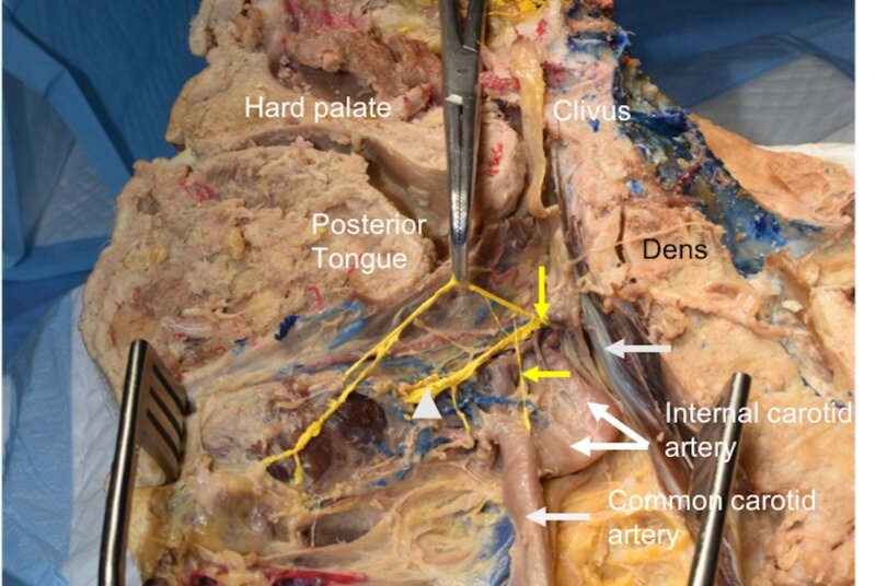
The carotid sinus nerve is known to convey baroreceptive fibers from the carotid sinus. Despite studies on the baroreflex pathway and the course and communications of the carotid sinus nerve with the surrounding nervous and vascular structures, there have been scant reports on variations in the origin of the carotid sinus nerve (CSN). We identified an unusual origin of the CSN. On the right side of a cadaveric specimen, the CSN was found to arise from two small rami extending from the external laryngeal nerve. Such a case can help better understand various pathways used to monitor the carotid sinus. Additionally, surgeries that manipulate the superior laryngeal nerve could possibly injure a variant carotid sinus nerve, as seen in the present case.

KEYWORDS:

cranial nerve ix; glossopharyngeal nerve; intercarotid plexus; vagus nerve

The Role of the Axillary Arch Variant in Neurovascular Syndrome of Brachial Plexus Compression.

Cureus. 2018 Jun 25;10(6):e2875. doi: 10.7759/cureus.2875.

Rai RIwanaga JLoukas MOskouian RJTubbs RS.


Abstract

Axillary arch muscles are often found. In their course through this area, they might interfere with regional neurovascular structures. This case report will examine the presence of the axillary arch muscle and its implication in brachial plexus compression. During routine dissection of the left axilla and upper limb, a variant muscle (axillary arch muscle) was identified arising from the distal tendon of the latissimus dorsi and extending laterally to insert onto the deep surface of the tendon of insertion of the deltoid muscle. In adduction of the upper limb, the muscle was lax without compression of any underlying neurovascular structures. However, in abduction, the aberrant band of muscles compressed the proximal branches of the brachial plexus. Clinicians should be aware of this anatomical variant and its clinical significance in neurovascular compression including brachial plexus compression, thoracic outlet syndrome, and hyperabduction syndrome. This literature will review the anatomy of the axillary arch and its clinical correlate regarding signs, symptoms, diagnosis, and treatment in brachial plexus compression.

KEYWORDS:

axillary arch; brachial plexus compression; langer’s arch; median nerve; neurovascular

A Variant of the Sciatic Nerve and its Clinical Implications.

Cureus. 2018 Jun 25;10(6):e2874. doi: 10.7759/cureus.2874.

Dupont GUnno FIwanaga JOskouian RJTubbs RS.


Abstract

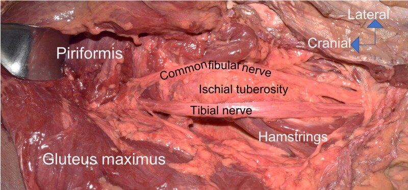
Variants of gluteal neural anatomy are important to consider, especially during surgical approaches to the hip. During the routine dissection of the gluteal region, a variant of the sciatic nerve was found where the nerve left the pelvis fully split into its tibial and common fibular components. Intrapelvically and extrapelvically, there was no splitting of the two components by the piriformis muscle. Distally, the two parts of the nerve were draped over the medial and lateral edges of the ischial tuberosity. To avoid iatrogenic injury to the sciatic nerve during invasive or surgical approaches to this region, all possible anatomical variations, such as the one presented herein, should be appreciated by the clinician.

KEYWORDS:

anatomy; gluteal region; iatrogenic; nerve injury; posterior thigh; surgery

Comprehensive Review of the Cardinal Ligament.

Cureus. 2018 Jun 20;10(6):e2846. doi: 10.7759/cureus.2846.

Eid SIwanaga JOskouian RJLoukas MTubbs RS.


Abstract

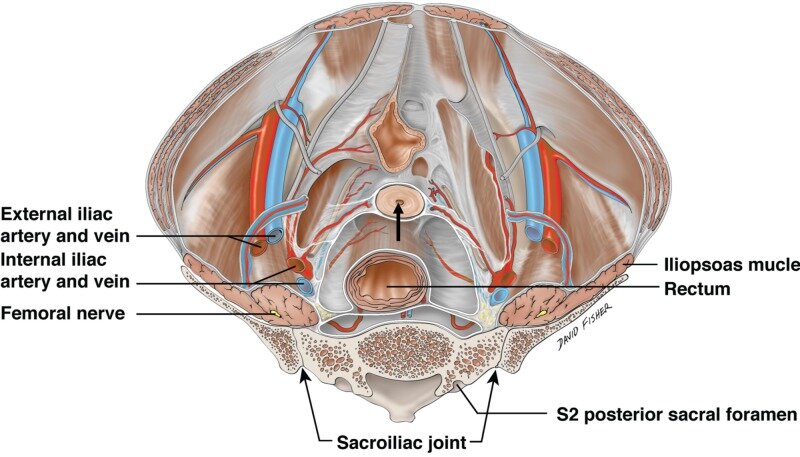
The cardinal ligament is thought to be one of the important structures in providing support and stabilization for the pelvic organs e.g., the uterus. However, many discrepancies exist in the literature regarding terminology, anatomy, and histology. The cardinal ligament attaches the lateral side of the vagina and cervix to the lateral pelvic wall, which provides support to the vagina and cervix. Studies have shown variable findings in the collagen content and distribution in the cardinal ligaments of women with a prolapsed uterus. Uterine vessels and the branches of the inferior hypogastric plexus travel in the cardinal ligament, which is of great importance during pelvic surgeries. Cervical cancer may lead to metastatic spread to the lymph nodes juxtaposed to the cardinal ligaments. This review aims to highlight the clinical significance and surgical importance of the cardinal ligament with a comparison with previous studies.

KEYWORDS:

cardinal ligament; parametrium; pelvic prolapse; transverse cervical ligament; uterus

The Deep Fascia of the Forearm and the Ulnar Nerve: An Anatomical Study.

Cureus. 2018 Jun 19;10(6):e2842. doi: 10.7759/cureus.2842.

Choi PJNwaogbe CIwanaga JGeorgiev GPOskouian RJTubbs RS.


Abstract

Introduction A reoperation for a cubital tunnel syndrome is not uncommon. Patients often complain of sensorimotor symptoms in the ulnar nerve distribution after their primary surgery. The documented etiologies for such a phenomenon include a "new" kinking of the distal ulnar nerve and a "new" compression of the ulnar nerve by the fascial septum in between or tendinous bands over the muscles of the forearm. The deep fascial plane along which the ulnar nerve travels in the forearm has had scant attention. We present an anatomical study to provide a better understanding of such etiologies to aid physicians in performing successful primary ulnar nerve release that does not lead to risky reoperations and ultimately yields improved patient satisfaction. Materials and methods The forearms of 12 fresh frozen cadavers (24 arms) underwent dissection, during which the fascial relationships between the ulnar nerve and muscles of the anterior compartment were explored with a blunt technique. The relationship between the fascial planes and the ulnar nerve was quantitatively and qualitatively documented. The ranges of motion of the elbow were also observed for any potential compression points on the nerve during the movement. Results In all specimens (n = 24), the ulnar nerve entered the forearm between the humeral and ulnar heads of the flexor carpi ulnaris, after which it routed deep to a deep fascia between the anterior surface of the flexor carpi ulnaris and the posterior surface of the flexor digitorum superficialis. Ulnar nerve branches to the flexor carpi ulnaris pierced this fascial septum while en route to the posterior surface of the muscle. Medially, the branches to the flexor digitorum profundus also pierced this fascial plane. In most arms, the fascia became thinner near the junction between the proximal two-thirds and distal one-third of the forearm. On no side was the ulnar nerve found to be grossly compressed by this deep fascia. However, with the extension of the elbow, a degree of angulation of the proximal ulnar nerve was observed due to its compact connection with the deep fascia. Conclusion Our study revealed that there is an intimate relationship between the ulnar nerve and the deep fascia of the forearm. Since the ulnar branches to the overlying flexor carpi ulnaris pierce this deep structure, a care should be given to its anatomical course during surgery in this region to prevent denervation of the muscle.

KEYWORDS:

flexor carpi ulnaris; nerve entrapment; recurrent cubital tunnel syndrome; revision surgery; ulnar nerve

Review of the Variations of the Superficial Veins of the Neck.

Cureus. 2018 Jun 18;10(6):e2826. doi: 10.7759/cureus.2826.

Dalip DIwanaga JLoukas MOskouian RJTubbs RS.


Abstract

The venous drainage of the neck can be characterized into superficial or deep. Superficial drainage refers to the venous drainage of the subcutaneous tissues, which are drained by the anterior and external jugular veins (EJVs). The brain, face, and neck structures are mainly drained by the internal jugular vein (IJV). The superficial veins are found deep to the platysma muscle while the deep veins are found encased in the carotid sheath. The junction of the retromandibular vein and the posterior auricular vein usually form the EJV, which continues along to drain into the subclavian vein. The anterior jugular vein is usually formed by the submandibular veins, travels downward anterior to the sternocleidomastoid muscle (SCM), and drains either into the EJV or the subclavian vein. Other superficial veins of the neck to consider are the superior, middle, and inferior thyroid veins. The superior thyroid and middle thyroid veins drain into the IJV whereas the inferior thyroid vein usually drains into the brachiocephalic veins.

KEYWORDS:

external jugular; internal jugular; superficial; thyroid vein; vein

Ophthalmic Artery Aneurysm in a Cadaver: Case Report.

Cureus. 2018 Jun 16;10(6):e2818. doi: 10.7759/cureus.2818.

Dalip DIwanaga JLoukas MOskouian RJTubbs RS.


Abstract

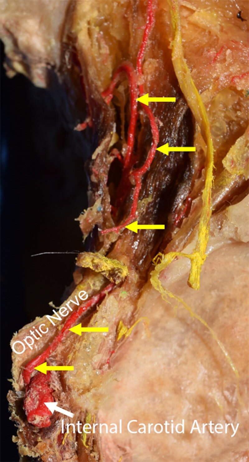
The ophthalmic artery arises from the supraclinoid segment of the internal carotid artery (ICA) and enters the orbit through the optic canal. It perfuses the orbit and the orbit apparatus. Ophthalmic artery aneurysms (OAA) account for 0.5% to 11% of all intracerebral aneurysms. Patients are usually asymptomatic but, in some cases, patients can present with ophthalmoplegia and total blindness if these aneurysms rupture. Aneurysms are usually diagnosed using computed tomography (CT) angiography but can also be seen on magnetic resonance imaging (MRI) and four-vessel digital subtraction angiography. Treatment of OAA entails either surgical or endovascular approaches with the mortality rate for surgical treatment as high as 25%, whereas embolization with balloon therapy is deemed safer with mortality rates around 9%. Recent techniques of embolization coiling have had even better results.

KEYWORDS:

aneurysm; embolization; ophthalmic artery; ophthalmoplegia

An Unusual Back Muscle Identified Bilaterally: Case Report.

Cureus. 2018 Jun 15;10(6):e2816. doi: 10.7759/cureus.2816.

Altafulla JJPatel MTubbs RSIwanaga JLitvack Z.


Abstract

Most muscular structures in the human body are named based on their function, origin/insertion, or shape. During routine dissection of the back, an unusual muscle was found deep to the rhomboid muscles. The details of this case and a review of the extant literature are provided.

KEYWORDS:

anomaly; myology; respiration; ribs; serratus posterior superior; variation

Pelvic Kidney: A Review of the Literature.

Cureus. 2018 Jun 9;10(6):e2775. doi: 10.7759/cureus.2775.

Eid SIwanaga JLoukas MOskouian RJTubbs RS.


Abstract

Kidney development is a complex process that begins during the sixth to eighth weeks of life. Failure of ascent of the kidney will cause the kidney to remain in the pelvis i.e., pelvic kidney. Here, we review this entity in detail and illustrate such embryological derailment. In most cases, a pelvic kidney is an incidental finding and is usually asymptomatic. Anatomic variations of the renal vasculature have been reported in cases of pelvic kidneys and these are highlighted in this review. Clinicians who treat patients for renal or pelvic disease or interpret images of the pelvis should be well informed of the anatomy and embryology of the pelvic kidney.

KEYWORDS:

anatomic variation; anatomy; ectopic kidney; horseshoe kidney; pelvic kidney