The Current Understanding of MicroRNA's Therapeutic, Diagnostic, and Prognostic Role in Chordomas: A Review of the Literature.

Cureus. 2018 Dec 24;10(12):e3772. doi: 10.7759/cureus.3772.

Choi PJOskouian RJTubbs RS.


Abstract

Chordomas are primary low-grade bone tumors derived from the embryonic notochord that make up less than 5% of all osseous malignancies and commonly affect the spine at its vertebral body and at its two ends i.e., skull base and the sacrum. Although histologically defined to be low-grade, chordoma is locally destructive, metastatic, and has a serious recurrence rate, which all contribute to the dismal median survival rate of six years. Its locally destructive nature places the adjacent vital neurovascular structures at risk, making an en-bloc resection a challenge. This tumor is also known to show high resistance to currently available chemoradiotherapy, although the benefit of proton beam therapy for skull base chordoma has been demonstrated. There is an additional need to focus our attention on investigating the molecular biology of this chemoradiotherapy-resistant tumor to develop a more targeted therapy, which has additional diagnostic and prognostic values. In this paper, we discuss the therapeutic, diagnostic, and prognostic role of microRNAs (miRNAs) in chordomas.

KEYWORDS:

biological target; chemoradiotherapy resistance; differential expression; microrna; mirna profiling; prognosis; sacral chordoma

First Report of a Bifid Mandibular Canal Containing a Large Vein Draining into the Anterior Jugular Vein.

Kurume Med J. 2018 Dec 21;65(1):27-30. doi: 10.2739/kurumemedj.MS651004. Epub 2018 Aug 30.

Iwanaga JWilson CSimonds EVetter MKusukawa JYamaki KIOskouian RJTubbs RS.


Abstract

The bifid mandibular canal (BMC) is an anatomical variant of the mandible that is often observed on cone-beam computed tomographic images. We identified a BMC during routine cadaveric dissection. The upper mandibular canal contained the inferior alveolar nerve and artery, and the lower mandibular canal contained a large inferior alveolar vein. This latter vein left the mandible through a lateral lingual foramen and joined the anterior jugular vein. Additionally, this vein gave rise to small tributaries to the mental foramen and anterior surface of the mandible from the second mandibular canal. To our knowledge, this is the first report illustrating the contents of a BMC and drainage of a vein into the large anterior jugular vein.

KEYWORDS:

Anatomy; anatomic variation; cadaver; inferior alveolar nerve; mandible; vein

Clinical Anatomy of Blockade of the Pterygopalatine Ganglion: Literature Review and Pictorial Tour Using Cadaveric Images.

Kurume Med J. 2018 Dec 21;65(1):1-5. doi: 10.2739/kurumemedj.MS651001. Epub 2018 Aug 30.

Iwanaga JWilson CSimonds EVetter MSchmidt CYilmaz EChoi PJOskouian RJTubbs RS.


Abstract

Pterygopalatine ganglion block (sphenopalatine ganglion block) is a well-known procedure for treating cluster headache and for relieving cancer pain. In this review, the history and anatomy of the pterygopalatine ganglion are discussed, and images, including computed tomography and endoscopy, are presented to improve understanding of the clinical anatomy of the ganglion regarding the block procedure.

KEYWORDS:

anatomy; cadaver; cluster headache; pterygopalatine ganglion block; sphenopalatine ganglion block

The Conchal Vascular Foramen of the Posterior Auricular Artery: Application to Conchal Cartilage Grafting.

Kurume Med J. 2018 Dec 21;65(1):7-10. doi: 10.2739/kurumemedj.MS651002. Epub 2018 Aug 30.

Wilson CIwanaga JSimonds EYilmaz EOskouian RJTubbs RS.


Abstract

The posterior auricular artery (PAA), a branch of the external carotid artery, gives rise to a conchal network formed by PAA perforators through the conchal floor of the auricle. However, this branch and its entrance (foramen) to the anterior concha, is rarely illustrated in the literature and has not been studied in detail. Therefore, we aimed to investigate the morphology of the perforating artery (PA) and its vascular foramen (VF). Ten sides from five formalin-fixed frozen Caucasian cadaveric heads were used. The number, diameter of the VF, diameter of the perforating artery (PA), shape of the VF (circular or oval), distance from the middle of the tragus and origin of the artery were documented. The number of VF ranged from 1 to 2; one was seen on 90% of the sides and two were seen on 10% of the sides. The VF was oval in 36% of the sides and circular in the remaining 64%. The mean diameter of the long and short axes of the VF, and PA was 2.0±1.4 mm, 1.3±0.9 mm, and 0.7±0.4 mm, respectively. Diameter of the PA was 1.0 mm or greater in 18% of the sides. The mean distance from the middle of the tragus to the VF was 10.7±2.6 mm. The perforating artery of the concha originated from the posterior auricular artery on all 11 sides.

KEYWORDS:

anatomy; cadaver; ear auricle; ear cartilage; external carotid artery

Cerebellar tonsil ectopia measurement in type I Chiari malformation patients show poor inter-operator reliability.

Abstract

BACKGROUND:

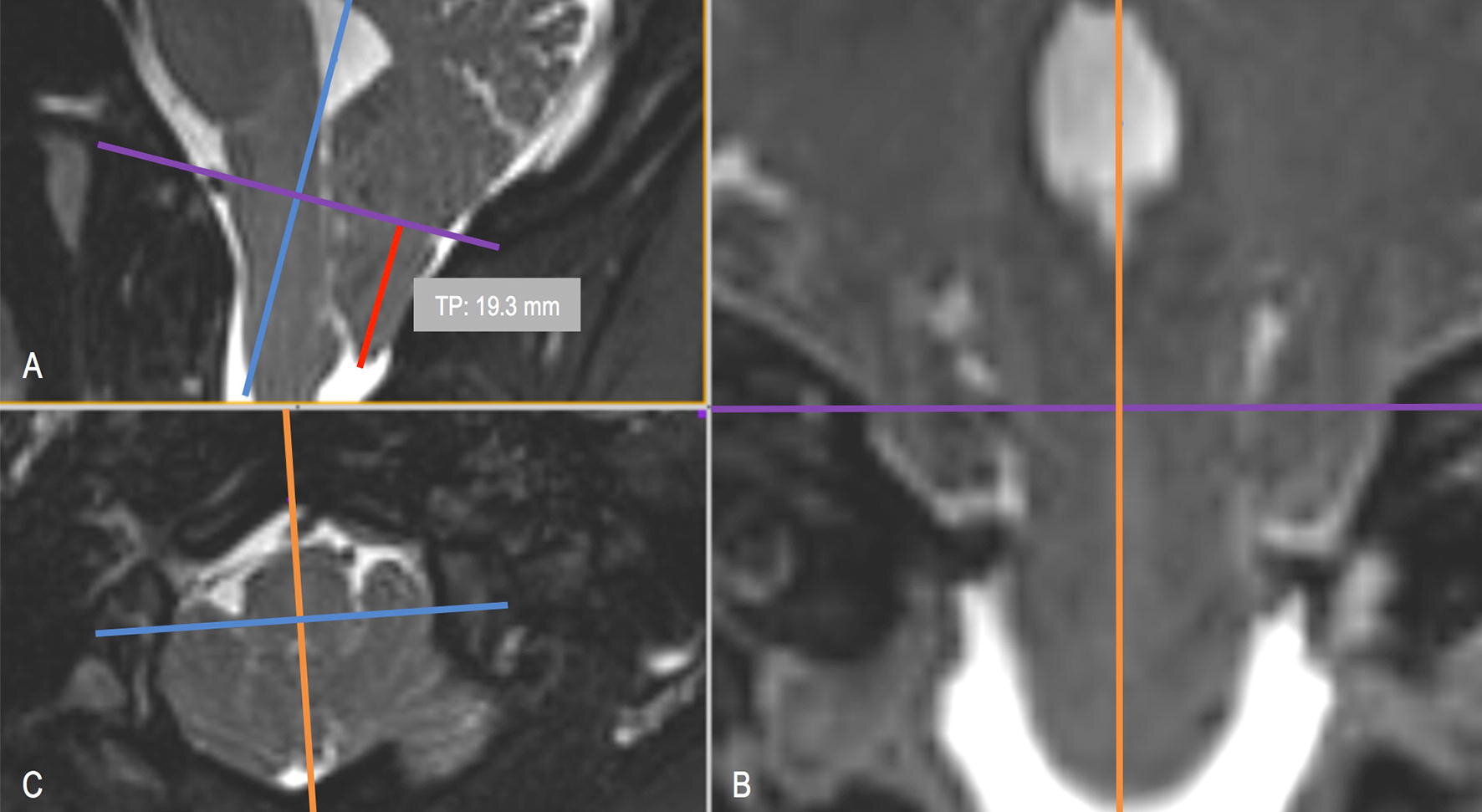
Type 1 Chiari malformation (CM-I) has been historically defined by cerebellar tonsillar position (TP) greater than 3-5 mm below the foramen magnum (FM). Often, the radiographic findings are highly variable, which may influence the clinical course and patient outcome. In this study, we evaluate the inter-operator reliability (reproducibility) of MRI-based measurement of TP in CM-I patients and healthy controls.

METHODS:

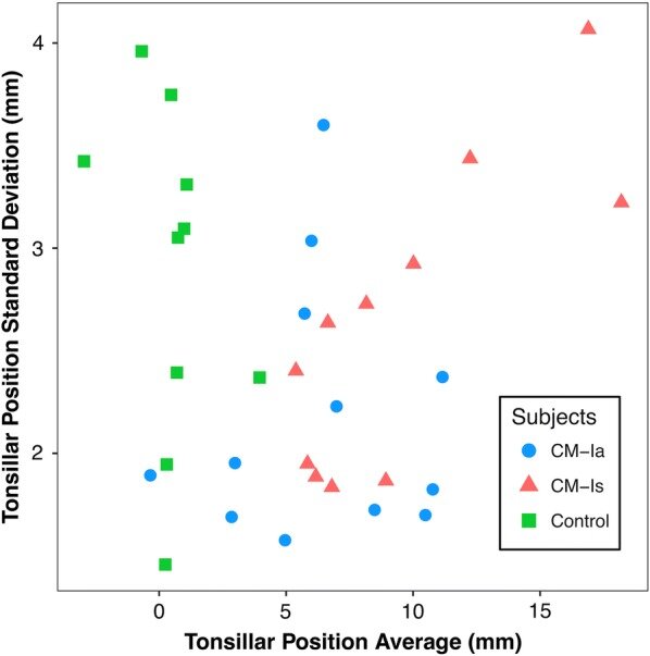
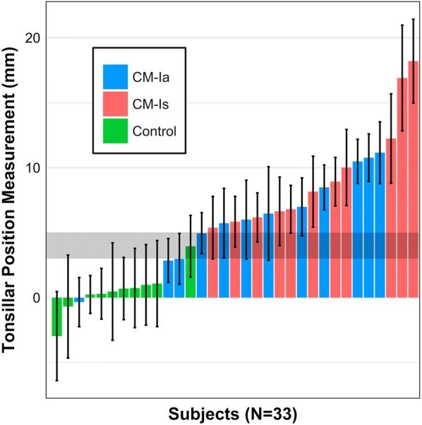
Thirty-three T2-weighted MRI sets were obtained for 23 CM-I patients (11 symptomatic and 12 asymptomatic) and 10 healthy controls. TP inferior to the FM was measured in the mid-sagittal plane by seven expert operators with reference to McRae's line. Overall agreement between the operators was quantified by intraclass correlation coefficient (ICC).

RESULTS:

The mean and standard deviation of cerebellar TP measurements for asymptomatic (CM-Ia) and symptomatic (CM-Is) patients in mid-sagittal plane was 6.38 ± 2.19 and 9.57 ± 2.63 mm, respectively. TP measurements for healthy controls was 0.48 ± 2.88 mm. The average range of TP measurements for all data sets analyzed was 7.7 mm. Overall operator agreement for TP measurements was relatively high with an ICC of 0.83.

CONCLUSION:

The results demonstrated a large average range (7.7 mm) of measurements among the seven expert operators and support that, if economically feasible, two radiologists should make independent measurements before radiologic diagnosis of CM-I and surgery is contemplated. In the future, an objective diagnostic parameter for CM-I that utilizes automated algorithms and results in smaller inter-operator variation may improve patient selection.

KEYWORDS:

Cerebellar tonsil; Inter-operator reliability; MRI; Morphometric; Syringomyelia; Type 1 Chiari malformation

Bilateral sternocleidomastoid variant with six distinct insertions along the superior nuchal line.

Anat Cell Biol. 2018 Dec;51(4):305-308. doi: 10.5115/acb.2018.51.4.305. Epub 2018 Dec 29.

Dupont GIwanaga JAltafulla JJLachkar SOskouian RJTubbs RS.


Abstract

Anatomical variations of the sternocleidomastoid muscle (SCM) have been observed to occupy multiple origins and insertion points and have supernumerary heads, sometimes varying in thickness. During routine dissection, a SCM was observed to have six distinct insertions that interface with the course of the superior nuchal line, ending at the midline, bilaterally. This variation was also seen to receive innervation from the accessory nerve as well as the great auricular nerve. To our knowledge, this variant of supernumerary insertions and nerve innervations has not yet been reported. These variants may pose as problematic during surgical approaches to the upper neck and occiput, and should thus be appreciated by the clinician. Herein we discuss the case report, possible embryological origins, and the clinical significance of the observed variant SCM.

KEYWORDS:

Anatomy; Mastoid; Neck surgery; Sternocleidomastoid; Variant

A comprehensive review of Mongolian spots with an update on atypical presentations.

Childs Nerv Syst. 2018 Dec;34(12):2371-2376. doi: 10.1007/s00381-018-3929-0. Epub 2018 Aug 6.

Alimi YIwanaga JLoukas MOskouian RJRizk EOakes WJTubbs RS.


Abstract

PURPOSE:

Mongolian spots (MS) are grayish-blue macules that typically present in the lumbosacral and gluteal regions of newborns. They are mostly benign and tend to resolve with age. This review brings to light recent findings and disorders associated MS.

METHODS:

In this paper, we review the literature, highlight recent cases and disorders associated with MS, and emphasize how newborns presenting with atypical MS should undergo appropriate screening.

RESULTS:

Atypical MS on other body parts are more likely to persist and might be an indication for further screening. Recent studies have shown persistent and extensive MS to be associated with various genetic disorders such as lysosomal storage diseases (LSDs) and phakomatosis pigmentovascularis.

CONCLUSION:

Physicians should be aware of atypical MS and related conditions in order to further assess these patients for risk of any underlying genetic disorders.

KEYWORDS:

Cleft lip; Dermal melanocytosis; Hurler’s syndrome; Lysosomal storage diseases; MPS 1; Mongolian spots

Atraumatic Vertebral Arteriovenous Fistula: A Rare Entity with Two Case Reports.

World Neurosurg. 2018 Dec;120:66-71. doi: 10.1016/j.wneu.2018.08.160. Epub 2018 Aug 30.

Rai RIwanaga JWang BPatel ABentley JLoh YMonteith STubbs RS.


Abstract

BACKGROUND:

A vertebral artery arteriovenous fistula is rare and usually due to trauma. Atraumatic cases are quite rare. We reported 2 cases and a review of other reported studies.

CASE DESCRIPTION:

A spontaneous vertebral-venous fistula is rare, and the 2 cases presented illustrate an underlying spontaneous etiology. The first patient presented with a spontaneous fistula, and the second case occurred in a patient with neurofibromatosis type 1. In both cases, the fistulas were diagnosed using computed tomography angiography and treated with occlusion via coil embolization.

CONCLUSIONS:

Vascular changes are known in patients with neurofibromatosis. A proposed pathogenesis of fistula is that the fragility and defective nature of the arterial wall could be a predisposing factor or it might be congenital. Understanding the clinical symptoms, diagnosis, and effective management strategies are important for physicians treating patients with a vertebral artery arteriovenous fistula.

Copyright © 2018. Published by Elsevier Inc.

KEYWORDS:

Fistula embolization; Neurofibromatosis; Spontaneous arteriovenous fistula; Vertebral artery

Dominique Jean Larrey (1766-1842) and His Contributions to Military Medicine and Early Neurosurgery.

World Neurosurg. 2018 Dec;120:96-99. doi: 10.1016/j.wneu.2018.08.159. Epub 2018 Aug 30.

Ramdhan RCRai RBrooks KNIwanaga JLoukas MTubbs RS.


Abstract

Dr. Dominique Jean Larrey was a pioneer in the field of military surgery. His creative innovation and drive to improve the quality of medical services available to those injured during war guided his achievements in medicine. Dr. Larrey has often been referred to as "the father of emergency medical services" and "the father of modern military medicine." His contributions to medicine continue to live on in common procedures and healthcare systems today, such as aspiration of pericardial effusion and drainage of hemothorax and empyema. Based on his own writings, he treated multiple cases of intracranial injury, making him an early, but often forgotten, pioneer of neurosurgery.

Copyright © 2018. Published by Elsevier Inc.

KEYWORDS:

Biography; Dominique Jean Larrey; Emergency medicine; History; Military medicine; Neurosurgery

Superior Hypogastric Plexus and Its Surgical Implications During Spine Surgery: A Review.

World Neurosurg. 2018 Dec;120:163-167. doi: 10.1016/j.wneu.2018.08.170. Epub 2018 Aug 30.

Eid SIwanaga JChapman JROskouian RJLoukas MTubbs RS.


Abstract

The superior hypogastric plexus (SHP) is a complex nervous collection located at the lumbosacral region below the level of the aortic bifurcation. As a part of the autonomic nervous system, it is an extension of the preaortic plexuses and continues bilaterally as the hypogastric nerves that ultimately contribute to the inferior hypogastric plexus. Although commonly described as a plexiform structure, several morphologic variations exist. Damage to the SHP can occur during anterior and anterolateral approaches to the lumbosacral spine leading to dysfunction of the abdominopelvic viscera. Visceral afferents travel in the SHP and are responsible for transmitting pain. Management therapies such as SHP blockade or presacral neurectomy can reduce pelvic pain caused by cancer and nonmalignant etiologies. This review highlights some of the recent findings regarding the nature of the SHP.

Copyright © 2018. Published by Elsevier Inc.

KEYWORDS:

Autonomic nervous system; Hypogastric nerves; Pelvic pain; Superior hypogastric plexus; Superior hypogastric plexus block

Wound Closure After Posterior Multi-level Lumbar Spine Surgery: An Anatomical Cadaver Study and Technical Note.

Cureus. 2018 Nov 14;10(11):e3595. doi: 10.7759/cureus.3595.

Yilmaz ETawfik TO'Lynnger TMIwanaga JBlecher RAbdul-Jabbar ATubbs RSSchmidt CKOskouian RJChapman J.


Abstract

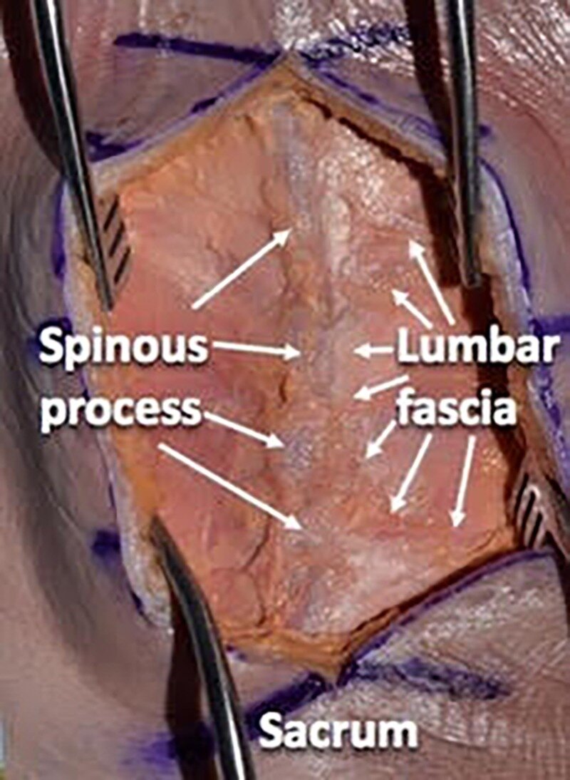
Meticulous attention to wound closure in posterior lumbar spine surgery is an important principle in reducing surgical site infections. We detail standardized wound closure used for posterior lumbar spine surgery at a tertiary care referral center and illustrate this as a step-by-step cadaveric dissection. The lumbar spine of a cadaveric specimen (male, 73 years at death) was used for dissection. Standardizing wound closure in posterior lumbar spine surgery may help limit wound complications and infection. Some key points of our technique, as demonstrated on a cadaveric specimen, include separating fascial compartments, avoiding suture abscesses, and creating a tension-free wound.

KEYWORDS:

fascia; lumbar; spine surgery; subcutaneous; technique; wound closure

Long-term impact of abusive head trauma in young children.

Child Abuse Negl. 2018 Nov;85:39-46. doi: 10.1016/j.chiabu.2018.08.011. Epub 2018 Aug 23.

Nuño MUgiliweneza BZepeda VAnderson JECoulter KMagana JNDrazin DBoakye M.


Abstract

OBJECTIVE:

Abusive head trauma is the leading cause of physical abuse deaths in children under the age of 5 and is associated with severe long-lasting health problems and developmental disabilities. This study evaluates the long-term impact of AHT and identifies factors associated with poor long-term outcomes (LTOs).

METHODS:

We used the Truven Health MarketScan Research Claims Database (2000-2015) to identify children diagnosed with AHT and follow them up until they turn 5. We identified the incidence of behavioral disorders, communication deficits, developmental delays, epilepsy, learning disorders, motor deficits, and visual impairment as our primary outcomes.

RESULTS:

The incidence of any disability was 72% (676/940) at 5 years post-injury. The rate of developmental delays was 47%, followed by 42% learning disorders, and 36% epilepsy. Additional disabilities included motor deficits (34%), behavioral disorders (30%), visual impairment (30%), and communication deficits (11%). Children covered by Medicaid experienced significantly greater long-term disability than cases with private insurance. In a propensity-matched cohort that differ primarily by insurance, the risk of behavioral disorders (RD 36%), learning disorders (RD 30%), developmental delays (RD 30%), epilepsy (RD 18%), and visual impairment (RD 12%) was significantly higher in children with Medicaid than kids with private insurance.

CONCLUSION:

AHT is associated with a significant long-term disability (72%). Children insured by Medicaid have a disproportionally higher risk of long-term disability. Efforts to identify and reduce barriers to health care access for children enrolled in Medicaid are critical for the improvement of outcomes and quality of life.

Copyright © 2018 Elsevier Ltd. All rights reserved.

KEYWORDS:

Abusive head trauma (AHT); Injury severity score (ICISS); Long-term outcomes; Propensity matched cohort; Risk difference (RD)

Incidence of Foramen Tympanicum (of Huschke): Comparing Cadaveric and Radiologic Studies.

J Craniofac Surg. 2018 Nov;29(8):2348-2352. doi: 10.1097/SCS.0000000000004784.

Pękala JRPękala PASatapathy BHenry BMSkinningsrud BPaziewski MTubbs RSTomaszewski KA.


Abstract

PURPOSE:

The foramen tympanicum (FT) represents a developmental anomaly that forms due to incomplete fusion of processes of the tympanic ring. Its presence in the population is controversial and it has been associated with a number of otologic complications. The aim of this study was to systematically analyze the prevalence, anatomical characteristics, and ethnic variations of the FT and compare these parameters in cadaveric and radiologic studies.

METHODS:

An extensive search was conducted through the major electronic databases, and identified articles were separated into 2 groups based on their methodology: cadaveric and radiologic studies. Data extracted included study modality, prevalence data, ethnicity, gender, side, laterality, and diameter.

RESULTS:

A total of 8 studies (n = 2671 patients) were included into our meta-analysis. The main findings revealed that the prevalence of the FT in the population is 14.9%, it is more often unilateral (62.5%) than bilateral (37.5%), it is most often reported in Asia (21.4%), and it is more prevalent in cadavers (21.2%) than in radiologic studies (8.8%) (not statistically significant).

CONCLUSION:

As the FT is present in more than one-tenth of the population, it is important to consider the possibilities of its presence when undertaking surgical procedures in the temporomandibular joint and ear region and plan accordingly to avoid injuries. Clinicians should keep it as one of the possible diagnoses while confronted with patients presenting with otologic complications. The use of newer imaging techniques was recommended, such as cone-beam computer tomography to detect the FT prior to surgery.

Guest UserComment
Pons ratio as a potential diagnostic biomarker for the detection of growth hormone deficiency in children.

Neurol India. 2018 Nov-Dec;66(6):1680-1684. doi: 10.4103/0028-3886.246236.

Tunçyürek ÖTurgut MÜnüvar TTubbs RSÖzsunar Y.


Abstract

INTRODUCTION:

Pituitary insufficiency (PI) needs further research to optimize treatment. Growth hormone deficiency (GHD) is a subtype of PI. The purpose of the present study is to investigate the possible relationship between GHD and the anatomical position of the pons as revealed by magnetic resonance imaging (MRI) in the pediatric age group. In the current study, we developed a novel and simple index using MRI that could provide an alternative to other indexes in the classical literature.

PATIENTS AND METHODS:

Brain MRI and clinical data of 48 children with suspected PI (22 females, 26 males; mean age 11.6 ± 2.2 years) were examined retrospectively. To estimate the location of the pons, the ratio of pons height over the axis between the dorsum sellae and the fourth ventricular hill in the sagittal plane (A) to the total height of the pons (B) was calculated (A/B). It was termed the pons ratio (PR). The PRs of children with or without a diagnosis of GHD were then compared statistically.

RESULTS:

Fifteen children were diagnosed with GHD and treated with growth hormone (GH), whereas the remaining 33 were reported normal. The mean PRs of the children diagnosed with GHD and given GH treatment (0.31 ± 0.07, range: 0.18-0.42) differed significantly from those without PI (0.26 ± 0.06, range: 0.17-0.44; P = 0.018).

CONCLUSION:

There appears to be an association between GHD and decreased PR in children receiving GH treatment. Spatial measurements of posterior fossa in radiological examinations may provide additional information that is helpful in the diagnosis of suspected cases of GHD.

KEYWORDS:

Magnetic resonance imaging; pediatric population; pituitary insufficiency; pons; posterior fossa

Type I Spinal Arteriovenous Fistula with Ventral Intradural Venous Drainage: A Proposal of a Modified Classification.

Asian J Neurosurg. 2018 Oct-Dec;13(4):1048-1052. doi: 10.4103/ajns.AJNS_100_17.

Adeeb NMoore JMAlturki AYBulsara KRGriessenauer CJPatel ASGupta RTubbs RSOgilvy CSThomas AJ.


Abstract

OBJECTIVES:

Spinal arteriovenous fistula (AVF) is the most common spinal vascular lesion and constitutes an abnormal communication between a feeder artery and a draining vein. Arterialization of the venous plexus leads to venous hypertension; consequent edema and congestion of the spinal cord are associated with progressive neurological decline.

PATIENTS AND METHODS:

In this report, we describe two unique cases of type I cervical spinal AVF, in which a radiculomeningeal artery forms an intradural fistula that drains into the ventral venous plexus.

RESULTS:

Both patients underwent surgical obliteration of the fistula with complete occlusion confirmed on postoperative angiography.

CONCLUSION:

Both cases do not fit into the current classification scheme. A modified classification is proposed.

KEYWORDS:

Arteriovenous; Type I; arteriovenous fistulas; classification; fistula; spinal; venous plexus; ventral

A novel marker for identifying and studying the membranes, barriers, and compartments surrounding peripheral nerves microscopically.

Clin Anat. 2018 Oct;31(7):1050-1057. doi: 10.1002/ca.23253. Epub 2018 Oct 26.

Reina MABoezaart APSala-Blanch XMonzó ETubbs RSServer ABigeleisen P.


Abstract

Recent anatomical discoveries indicate the importance of identifying membranes and compartments surrounding peripheral nerves into which local anesthetic agents can be injected and continuous nerve block catheters placed during regional anesthetic procedures. However, current markers used in anatomical studies have multiple drawbacks, specifically extravasation into noninjected locations, which can result in inadequate treatment. We studied a readily-available new marker, heparinized blood solution (HBS), which is easy to identify by microscopy and can remain in the nerve compartment into which it is deposited without distorting the tissue. We collected blood from 22 patients and prepared it as HBS. This was then injected into four fresh cadavers as in routine clinical practice for ultrasound-guided nerve blocks to form a so-called "doughnut" by "hydro-dissecting" at 32 sites. All samples, including nerves and neighboring tissues, were then prepared and examined by light microscopy. Although no deliberate intraneural injection was attempted, the marker was identified inside all the nerve compartments except the fascicles. Apart from leaking through the needle entry site in some instances, there was no extravasation of the HBS into neighboring nerve compartments in either direction. The tissues were not distorted and the erythrocytes did not form a thrombus. Nerve membranes and compartments could be clearly identified with routine staining. This technique enabled us to study the longitudinal and circumferential spread in all nerve compartments and to collect data for better interpretation of factors influencing an anesthetic nerve block and situations in which complications could possibly arise. HBS seemed superior to other markers because it did not leave the compartments into which it had been injected, did not distort the tissue, and was easily visible under the light microscope. Clin. Anat., 31:1050-1057, 2018.

© 2018 Wiley Periodicals, Inc.

KEYWORDS:

endoneurium; epineurium; nerve blockade; perineurium; peripheral nerve injuries

The clinical anatomy of dyspareunia: A review.

Clin Anat. 2018 Oct;31(7):1013-1017. doi: 10.1002/ca.23250. Epub 2018 Oct 26.

Alimi YIwanaga JOskouian RJLoukas MTubbs RS.


Abstract

Dyspareunia can be described as continuous unremitting or intermittent pain associated with intercourse. It can be classified based on the location of the pain - entry or deep dyspareunia, or based on when the pain was first experienced - primary or secondary dyspareunia. There are different causes of dyspareunia and some of the most important causes include the following: vulvodynia, postpartum dyspareunia, endometriosis, inadequate vaginal lubrication or arousal, and other anogenital causes such as hemorrhoids and anal fissures. In this review, our objective is to apply the anatomical knowledge of dyspareunia to patient care, increase awareness among clinicians about the diverse etiology of dyspareunia and ensure that the whole patient, not just the pain of dyspareunia is being treated as the causes of dyspareunia can be due to various pathologies. Clin. Anat., 31:1013-1017, 2018.

© 2018 Wiley Periodicals, Inc.

KEYWORDS:

anatomy; coital pain; deep dyspareunia; dyspareunia; entry dyspareunia; vulvodynia

Histological verification of atherosclerosis due to bends and bifurcations in carotid arteries predicted by hemodynamic model.

J Vasc Bras. 2018 Oct-Dec;17(4):280-289. doi: 10.1590/1677-5449.004118.

Singh RTubbs RS.


Abstract

BACKGROUND:

Tortuosity and bifurcations in carotid arteries alter the blood flow, causing atherosclerosis.

OBJECTIVES:

The aim of the present study is to analyze the effect of variant vascular anatomy in the cervical region on development of atherosclerosis by microanatomical examination.

METHODS:

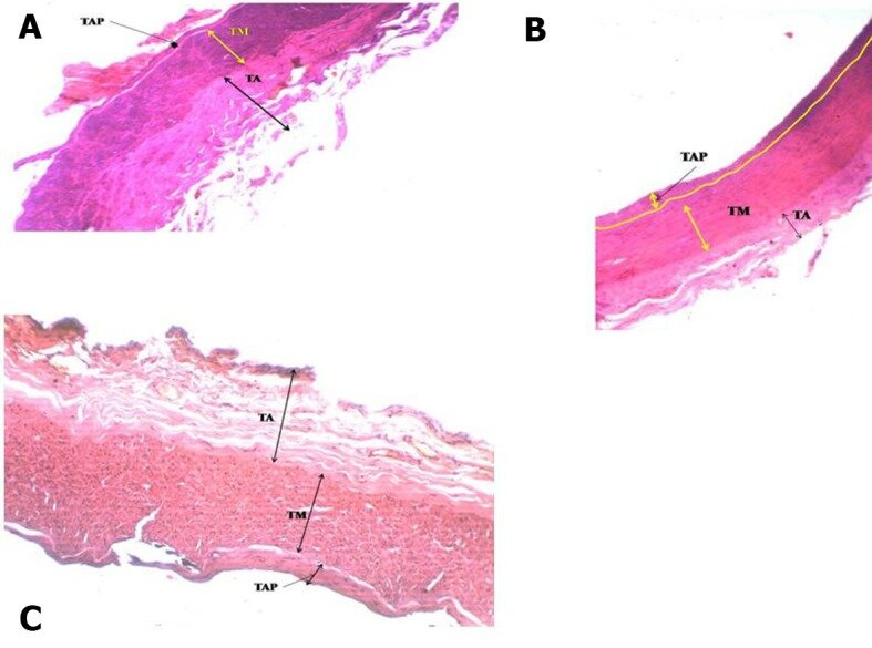
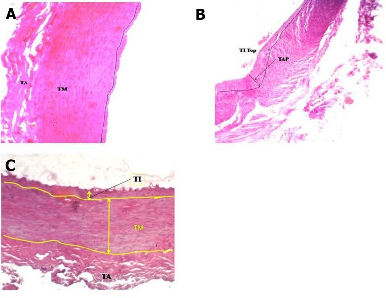
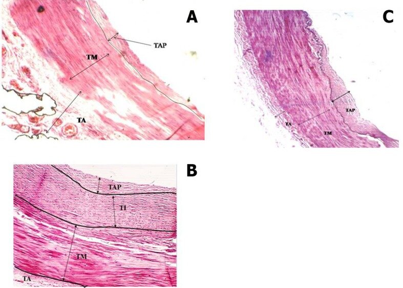
The effect of blood flow at anomalous bends and bifurcations was observed in right carotid arteries of a seventy year old female cadaver. Fifteen histological slides were prepared from the carotid arteries and interpreted to verify predictions of atherosclerosis.

RESULTS:

The model predicts atherosclerosis at bends, bifurcations and large aperture arteries. Microanatomical examination revealed presence of atherosclerosis of varying thickness at the bends and bifurcation in the right carotid arteries, as predicted. Atherosclerosis was also detected in the straight part of the wider common carotid artery. No atherosclerosis was observed in the contralateral carotid arteries. The variant carotid vascular anatomy consisting of bends, bifurcations and wider arteries revealed that the shear stress and velocity of blood flow are reduced at these anomalous sites.

CONCLUSIONS:

Anatomical anomalies such as bends and branching in the carotid arteries alter the irrigation pattern and generate biomechanical forces that cause turbulent flow and reduce shear stress/blood flow velocity. Decreased shear stress and velocity causes development of atherosclerosis. Histological slides established the presence of atherosclerosis at bends and bifurcations and in wider arteries.

KEYWORDS:

bifurcations; hemodynamic model; microanatomy; tortuosity

Arcuate Foramen: Anatomy, Embryology, Nomenclature, Pathology, and Surgical Considerations.

World Neurosurg. 2018 Oct;118:197-202. doi: 10.1016/j.wneu.2018.07.038. Epub 2018 Jul 17.

Ahn JDuran MSyldort SRizvi AD'Antoni AVJohal JIwanaga JOskouian RJTubbs RS.


Abstract

BACKGROUND:

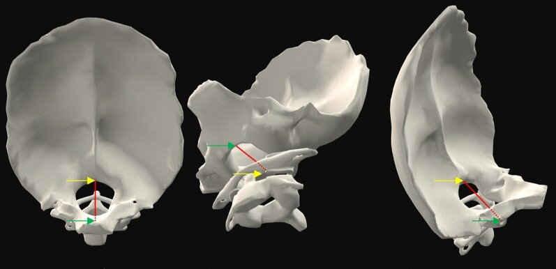
The arcuate foramen is an anatomic variant that is thought to arise from ossification of the posterior atlanto-occipital membrane. Owing to potential entrapment of the vertebral artery segment that traverses the foramen, vertebrobasilar ischemia may occur, and the person may experience vertigo, headache, or neck pain.

METHODS:

We reviewed the literature regarding anatomy (both human and comparative), embryology, nomenclature, pathology, and surgery of the arcuate foramen.

RESULTS:

Surgically, the presence of an arcuate foramen is important when placing screws into lateral masses of the atlas. In these cases, the screws can damage the V3 segment of the vertebral artery and/or the suboccipital nerve.

CONCLUSIONS:

It is important to review the current literature on the arcuate foramen to further understand its morphology and clinical relevance.

Copyright © 2018 Elsevier Inc. All rights reserved.

KEYWORDS:

Arcuate foramen; Atlas; Complications; Surgery; Vertebral artery