Recent Increase in the Rate of Spinal Infections May be Related to Growing Substance-use Disorder in the State of Washington

Spine (Phila Pa 1976). 2019 Feb 15;44(4):291-297. doi: 10.1097/BRS.0000000000002819.

Blecher RYilmaz EIshak BDrazin DOskouian RJChapman JR.


Abstract

OBJECTIVE:

The aim of this study was to evaluate trends in the incidence of spinal infections (SI) and the possible role of substance use disorder (SUD) as a key associated factor.

SUMMARY OF BACKGROUND DATA:

SI pose major diagnostic and therapeutic challenge in developed countries, resulting in substantial morbidity and mortality. With an estimated incidence of up to 1:20,000, recent clinical experiences suggest that this rate may be rising.

METHODS:

To evaluate a possible change in trend in the proportion of SI, we searched the Washington state Comprehensive Hospital Abstract Reporting System (CHARS) data during a period of 15 years. We retrieved ICD-9 and 10 codes, searching for all conditions that are regarded as SI (discitis, osteomyelitis, and intraspinal abscess), as well as major known SI-related risk factors.

RESULTS:

We found that the proportion of SI among discharged patients had increased by around 40% during the past 6 years, starting at 2012 and increasing steadily thereafter. Analysis of SI-related risk factors within the group of SI revealed that proportion of SUD and malnutrition had undergone the most substantial change, with the former increasing >3-fold during the same period.

CONCLUSION:

Growing rates of drug abuse, drug dependence, and malnutrition throughout the State of Washington may trigger a substantial increase in the incidence of spinal infections in discharged patients. These findings may provide important insights in planning prevention strategies on a broader level.

Guest UserComment
Sudden Headache and Blindness Due to Pituitary (Adenoma) Infarction: A Case Report.

Cureus. 2019 Feb 13;11(2):e4059. doi: 10.7759/cureus.4059.

Dupont GLachkar SIwanaga JTubbs RSIshak B.


Abstract

Pituitary infarction (PI) is a rare medical emergency appearing in patients with pituitary adenoma, presenting with sudden onset of severe headache, and often associated with vomiting, nausea, visual deterioration, and decreased consciousness. We describe an 83-year-old woman who presented with blindness after a history of severe headache. An urgent computed tomography (CT) scan of the brain had been arranged, and a massive pituitary adenoma with hemorrhage was observed, and admission to the neurosurgical department followed. A multidisciplinary team comprising a neurosurgeon, an ophthalmologist, and an endocrinologist is required to manage such cases. To confirm the diagnosis, an urgent magnetic resonance imaging (MRI) must be performed; CT scan can be indicated if MRI is contraindicated or not possible.

KEYWORDS:

opthalmoplegia; pituitary failure; pituitary infarction; sudden blindness

Pfeiffer type 2 syndrome: review with updates on its genetics and molecular biology.

Childs Nerv Syst. 2019 Feb 11. doi: 10.1007/s00381-019-04082-7. [Epub ahead of print]

Rai RIwanaga JDupont GOskouian RJLoukas MOakes WJTubbs RS.

Abstract

INTRODUCTION:

Pfeiffer syndrome is a rare autosomal dominant inherited disorder associated with craniosynostosis, midfacial hypoplasia, and broad thumbs and toes. The syndrome has been divided into three clinical subtypes based on clinical findings.

METHODS:

This review will specifically examine the most severe type, Pfeiffer syndrome type 2, focusing on its genetics and molecular biology.

CONCLUSION:

This subtype of the syndrome is caused by de novo sporadic mutations, the majority of which occur in the fibroblast growth factor receptor type 1 and 2 (FGFR1/2) genes. There is not one specific mutation, however. This disorder is genetically heterogeneous and may have varying phenotypic expressions that in various cases have overlapped with other similar craniosynostoses. A specific missense mutation of FGFR2 causing both Pfeiffer and Crouzon syndromes has been identified, with findings suggesting that gene expression may be affected by polymorphism within the same gene. Compared to other craniosynostosis-related disorders, Pfeiffer syndrome is the most extreme phenotype, as the underlying mutations cause wider effects on the secondary and tertiary protein structures and exhibit harsher clinical findings.

KEYWORDS:

Acrocephalosyndactyly; Cloverleaf skull; Craniosynostosis; Fibroblast growth factor receptor (FGFR); Pfeiffer syndrome

Ventral and dorsal tethering bands of the spinal cord in the same patient: a case report.

Childs Nerv Syst. 2019 Feb;35(2):389-391. doi: 10.1007/s00381-018-3993-5. Epub 2018 Oct 18.

Eid SIwanaga JLoukas MJerry Oakes WShane Tubbs R.


Abstract

Fibrous bands are one of the causes of tethered cord syndrome and these can be located on the dorsal or more rarely, ventral aspect of the dura mater. We report a case of dorsal and ventral tethering bands in the same patient found at surgery for tethered cord syndrome. Such fibrous bands restrict normal movement of the spinal cord and lead to a variety of neurological symptoms. To our knowledge, we report the first case of dorsal and ventral tethering bands of the spinal cord in the same patient. This case report aims to increase awareness of such anatomical variations and emphasize the importance of meticulous surgical dissection in order to identify such bands.

KEYWORDS:

De-tethering; Dorsal tethering; Meningocoele manqué; Spinal cord; Ventral tethering

FokI as a genetic factor of intervertebral disc degeneration: A PRISMA-compliant systematic review of overlapping meta-analyses.

J Clin Neurosci. 2019 Feb;60:36-43. doi: 10.1016/j.jocn.2018.09.028. Epub 2018 Oct 9.

Pekala PAHenry BMTaterra DPiwowar MVikse JTubbs RSTomaszewski KA.


Abstract

The association of FokI (rs2228570), a polymorphism of the vitamin D receptor gene, with intervertebral disc degeneration (IDD) has been investigated in a multitude of studies. However, conflicting results of these studies led to emergence of several meta-analyses over the past few years. Despite the increased statistical power, these meta-analyses have failed to provide uniform and conclusive data on the relationship of FokI with IDD. The aim of this study was to present a comprehensive review based on the most up-to-date meta-analyses on the association of FokI with IDD. A comprehensive search of all major databases was conducted to identify meta-analyses investigating relation between FokI and IDD. No date or language restrictions were applied. The Jadad decision algorithm was utilized to evaluate included meta-analyses and identify the one providing the best evidence. A total of 7 meta-analyses (n = 2580 original patients), that included six to ten case control studies, analyzed the association of FokI polymorphism with IDD. The meta-analysis of the highest quality supported the notion that overall there is no statistically significant association between FokI polymorphism and IDD. However, the authors showed that Caucasians have a reduced risk of IDD and Hispanics have an increased risk of IDD in the dominant and dominant/homozygous/heterozygous models of FokI polymorphism. While currently there is no evidence of an association between FokI polymorphism and IDD in the general population, ethnic predisposition has been shown.

Copyright © 2018 Elsevier Ltd. All rights reserved.

KEYWORDS:

FokI; Intervertebral disc degeneration; Vitamin D receptor; rs2228570

Prevalence and anatomy of the axillary arch and its implications in surgical practice: A meta-analysis.

Surgeon. 2019 Feb;17(1):43-51. doi: 10.1016/j.surge.2018.04.003. Epub 2018 May 22.

Taterra DHenry BMZarzecki MPSanna BPękala PACirocchi RWalocha JATubbs RSTomaszewski KA.


Abstract

PURPOSE:

The following research aimed to investigate the prevalence and anatomical features of the axillary arch (AA) - a muscular, tendinous or musculotendinous slip arising from the latissimus dorsi and that terminates in various structures around the shoulder girdle. The AA may complicate axillary lymph node biopsy or breast reconstruction surgery and may cause thoracic outlet syndrome.

METHODS:

Major electronic databases were thoroughly searched for studies on the AA and its variations. Data regarding the prevalence, morphology, laterality, origin, insertion and innervation of the AA was extracted and included in this meta-analysis. The AQUA tool was used in order to assess potential risk of bias within the included studies.

RESULTS:

The AA was reported in 29 studies (10,222 axillas), and its pooled prevalence estimate in this meta-analysis was found to be 5.3% of the axillas: unilaterally (61.6%) and bilaterally (38.4%). It was predominantly muscular (55.1% of the patients with the AA), originated from the latissimus dorsi muscle or tendon (87.3% of the patients with the AA), inserted into the pectoralis major muscle or fascia (35.2% of the patients with the AA), and was most commonly innervated by the thoracodorsal nerve (39.9% of the patients with the AA).

CONCLUSION:

The AA is a relatively common variant, hence it should not be neglected. Oncologists and surgeons should consider this variant while diagnosing an unknown palpable mass in the axilla, as the arch might mimic a neoplasm or enlarged lymph nodes.

Copyright © 2018 Royal College of Surgeons of Edinburgh (Scottish charity number SC005317) and Royal College of Surgeons in Ireland. Published by Elsevier Ltd. All rights reserved.

KEYWORDS:

Anatomy; Axilla; Axillary arch; Langer's axillary arch; Meta-analysis

A Novel Microsurgical Procedure for Revascularization of the Vertebral Artery.

World Neurosurg. 2019 Feb;122:e302-e306. doi: 10.1016/j.wneu.2018.10.026. Epub 2018 Oct 12.

Vetter MIwanaga JChoi PJYilmaz EOskouian RJTubbs RS.


Abstract

OBJECTIVE:

A broad armamentarium of microsurgical techniques affords flexibility to surgeons when choosing a procedure that is best tailored to fit the anatomy of an individual. Herein, we report on the feasibility of using the deep cervical artery (DCA) to revascularize the vertebral artery (VA) via a DCA-V3 bypass graft.

METHODS:

Fourteen DCAs from 7 injected cadaveric heads were located and traced. The diameter of the main trunk of the DCA was measured bilaterally at the C3 level. The proximal vertebral branches of each DCA were then severed and the main trunk of the DCA was transposed superiorly onto the V3 segment of the VA, which was also exposed bilaterally.

RESULTS:

The DCA was identified and traced bilaterally on all specimens. The diameter of the main trunk of the DCA at the C3 level ranged from 1.03 to 2.79 mm. The mean diameter of the main trunk of the DCA at this level was found to be 1.52 ± 0.60 mm for the right side and 1.46 ± 0.54 mm for the left side. After releasing the proximal vertebral branches of the DCA, all arteries were able to be transposed to the ipsilateral VA.

CONCLUSIONS:

Based on the mean diameter of the DCA reported in extant literature and this study, the blood flow volume of the DCA makes it a viable candidate to bypass the proximal VA.

Copyright © 2018 Elsevier Inc. All rights reserved.

KEYWORDS:

Anatomy; Bypass graft; Deep cervical artery; Posterior cervical triangle; Subclavian artery; Vertebral artery

Gerber's Ligament-A Forgotten Structure of the Craniocervical Junction.

World Neurosurg. 2019 Jan 17. pii: S1878-8750(19)30074-9. doi: 10.1016/j.wneu.2018.12.198. [Epub ahead of print]

Ishak BGnanadev RDupont GKikuta SAltafulla JIwanaga JTubbs RS


Abstract

BACKGROUND:

Ligaments of the craniocervical junction play a critical role in stabilizing this region. Gerber's ligament has more or less been forgotten and, to our knowledge, never studied.

METHODS:

Dissection of the craniocervical junction was performed in 15 fresh frozen cadavers. In the prone position, the posterior elements of the upper cervical vertebrae and occiput were removed. After removing the contents of the spinal canal and posterior cranial fossa, the dura mater and tectorial membrane were reflected. The superior band of the cruciform ligament was reflected. When Gerber's ligament was identified, its attachments and morphometry were recorded. Lastly, Gerber's ligament was observed while range of motion of the craniocervical junction was performed.

RESULTS:

Gerber's ligament was identified in 7 specimens (46.7%). This structure arose anteriorly from the junction of the superior band and transverse part of the cruciform ligament. Gerber's ligament was always found to be just deep to the superior band of the cruciform ligament but traveled more anteriorly to attach onto the posterior aspect of the dens approaching, but not attaching onto, its apex. Mean length was 11 mm, and mean width was 7 mm. Thickness of the ligament was 0.5-1.1 mm. The ligament was found to become taut with minimal rotation of the atlantoaxial joint and extension of the craniocervical junction.

CONCLUSIONS:

A good understanding of all ligaments of the craniocervical junction is important to surgeons and physicians treating patients with injury to the upper cervical spine.

Copyright © 2019 Elsevier Inc. All rights reserved.

KEYWORDS:

Accessory band; Anatomy; Craniocervical junction; Cruciform ligament; Transverse ligament

The Carotid Sinus Nerve and the First English Translation of Hering's Original Research on this Nerve.

Cureus. 2019 Jan 16;11(1):e3898. doi: 10.7759/cureus.3898.

Shoja MMRai RLachkar SIboroma Akobo SYilmaz ELoukas MBinello EGorjaian MGriessenauer CJIwanaga JTubbs RS.


Abstract

This paper provides a brief depiction of the life and achievements of the most iconic experiments of Heinrich Ewald Hering. The authors herein have presented a translation of his paper on the carotid sinus nerve in English; the original paper by Heinrich Ewald Hering, titled "Ueber die Wand des Sinus caroticus als Reizempfänger und den Sinusnerv als zentripetale Bahn für die Sinusreflexe" (1924), provides a detailed account of his experimental process and findings. He recognized that the sinus reflexes are mediated by a branch of the glossopharyngeal nerve (CN IX).

KEYWORDS:

baroreceptors; carotid sinus nerve; glossopharyngeal nerve; historical; vagus nerve

Anatomico-radiological Study of the Bifurcate Ligament of the Foot with Clinical Significance.

Cureus. 2019 Jan 8;11(1):e3847. doi: 10.7759/cureus.3847.

Kafka RMAveytua ILChoi PJDiLandro ACTubbs RSLoukas MMintz DNBaidya RKumar SSangari SKMtui EPD'Antoni AV.


Abstract

Introduction Lateral ankle sprain caused by forcible plantar flexion and inversion of the foot commonly damages the anterior talofibular ligament and other ligaments. Unfortunately, involvement of the bifurcate ligament (BL) is often overlooked when assessing such injuries in clinical practice and identification of this ligament on magnetic resonance (MR) scans can be challenging. Anatomically, the BL is a Y-shaped structure with two bands: the calcaneonavicular ligament (CNL) and calcaneocuboid ligament (CCL). There are few anatomical studies on the morphometric characteristics of the BL and even fewer biomechanical studies. Therefore, the objective of this anatomico-radiological study was to investigate the morphology of the BL using a multifaceted approach, and classify the fiber characteristics of the CNL and CCL. Materials and methods We measured the length and the width of 53 embalmed cadaveric feet. Meticulous dissection of each foot was performed to expose the BL. Measurements of the length, width, thickness, and shape of the CNL and CCL were taken using a digital caliper. We also documented the fiber orientation of each ligament, and used a goniometer to measure the bifurcation angle between the CNL and CCL via two methods. Confirmatory histologic analysis of the ligaments was performed and digital radiographs of the ligaments with attached radiopaque monofilament were taken. We also included an MR scan of the BL. Using descriptive and inferential statistics, we documented any significant relationships between the variables. Results  Mean (range) age at death of cadavers was 76 (42-94) years. The CNL was found in all the feet and the CCL was not present in 9.4% of the feet. Mean (standard deviation) length of the CNL and CCL was 22.7 (4.12) mm and 10.9 (2.53) mm, respectively. Mean (standard deviation) thickness of the CNL and CCL was 3.23 (1.56) mm and 1.48 (0.71) mm, respectively. Related to ligament morphology, the CNL was most frequently cord shaped (67.92%) and the CCL was most frequently flat shaped (83.33%). The mean bifurcation angle measured 32.75o and 29.31o in methods 1 and 2, respectively. The correlation between the two measured angles was very strong (p < 0.001). Discussion We found that 90.6% of feet had both the CNL and CCL, 9.4% had the CNL and no CCL, and none (0%) had the CCL and no CNL. These frequencies are similar to a recent Japanese study. Our sample of donors were American and predominantly white. Whether the difference in frequencies between the studies is related to ethnicity is unknown and requires future investigation. Interestingly, on average the CNLs were twice as long and twice as thick as the CCLs. The CCLs tended to be wider distally and tapered compared to the CNLs. Conclusions Our findings better classify the morphology and fiber orientation of the BL. Coupled with the radiographs and MR scan, our data may be of particular value to radiologists and surgeons. Our BL fiber orientation classification system and angle measurements can pave the way for future biomechanical studies to investigate any relationships between fiber type, angle, and strength of the constituent bands. More accurate descriptions of the BL should lead to improved diagnosis and treatment of ligamentous injuries of the foot.

KEYWORDS:

anatomy; ankle sprain; bifurcate ligament; calcaneocuboid ligament; calcaneonavicular ligament; foot; foot joints; orthopedic surgery; podiatry; radiology

Untreated incomplete isolated cleft palate: cadaveric findings.

Anat Sci Int. 2019 Jan;94(1):154-157. doi: 10.1007/s12565-018-00471-1. Epub 2018 Nov 30.

Kikuta SIwanaga J5, Kusukawa JOskouian RJTubbs RSShane Tubbs R.

Abstract

Isolated cleft palate without cleft lip is a rare deformity. Cleft lip and cleft palate can sometimes develop in combination with a syndrome due to genetic causes. Affected patients have morbidity through life from birth and experience comprehensive treatment for such clefts including surgery. It is extremely rare that the untreated clefts are found during routine cadaveric dissection, since many patients have treatment for clefts in developed countries. Herein, we present a case of an untreated incomplete isolated cleft palate on the right side of the maxilla. An oronasal fistula was found in the same location as the missing right maxillary lateral incisor, and the soft palate was considerably intruding into the hard palate but without penetration into the nasal cavity. The right incisivus labii superioris muscle forming the oral vestibule was incompletely torn with two fistulae. An incomplete bony defect was found on the right maxilla without oronasal or oroantral fistula. This paper may contribute to evaluating the disturbed site of the incomplete isolated cleft palate with no treatment.

KEYWORDS:

Anatomy; Cadaver; Deformity; Isolated cleft palate; Surgery

Clinical anatomy of pelvic pain in women: A Gynecological Perspective.

Clin Anat. 2019 Jan;32(1):151-155. doi: 10.1002/ca.23270. Epub 2018 Dec 3.

Eid SLoukas MTubbs RS.


Abstract

Pelvic pain is a common symptom that affects women worldwide and usually presents with variable range of severity, duration, and location. Several gynecological conditions may result in pelvic pain, and may have a variable presentation among patients. Pelvic pain creates a significant challenge and frustration to both patients and clinicians, which also creates a significant burden on the economy. It is necessary to tailor the management of pelvic pain to each individual patient to achieve optimal outcomes. Endometriosis, adenomyosis, and fibroids result in pelvic pain and may occur simultaneously. This review highlights some of the common gynecological etiologies of pelvic pain with a focus on anatomy, diagnosis, and management. Clin. Anat. 9999:1-5, 2018.

© 2018 Wiley Periodicals, Inc.

KEYWORDS:

abdominopelvic; adenomyosis; endometriosis; female; fibroids; gynecology; leiomyoma; ovarian remnant syndrome; pelvic pain

Our current understanding of the lymphatics of the brain and spinal cord.

Clin Anat. 2019 Jan;32(1):117-121. doi: 10.1002/ca.23308.

Dupont GSchmidt CYilmaz EOskouian RJMacchi Vde Caro RTubbs RS.


Abstract

The lymphatic system, segregated from the blood vascular system, is an essential anatomical route along which interstitial fluid, solutes, lipids, immune cells, and cellular debris, are conveyed. However, the way these mechanisms operate within the cranial compartment is mostly unknown. Herein, we review current understanding of the meningeal lymphatics, described anatomically over a century ago yet still poorly understood from a functional standpoint. We will delineate the cellular mechanisms by which the meningeal lymphatics are formed and discuss their unique anatomy. Furthermore, this review will discuss the recently-coined "glymphatic system" and the manner by which cerebrospinal fluid (CSF) and interstitial fluid (ISF) are exchanged and thus drained by the meningeal lymphatic vasculature as a key route for conveying cellular waste, solutes, and immune traffic to the deep cervical lymph nodes. The clinical relevance of the meningeal lymphatics will also be described, as they are relevant to various common defects of the lymphatic system. Clin. Anat. 32:117-121, 2019.

© 2018 Wiley Periodicals, Inc.

KEYWORDS:

CNS lymphatics; Prox1; VEGF-C; VEGFR-3; glymphatic system; meningeal lymphatics

An evidence-based approach to learning clinical anatomy: A guide for medical students, educators, and administrators.

Clin Anat. 2019 Jan;32(1):156-163. doi: 10.1002/ca.23298. Epub 2018 Nov 26.

D'Antoni AVMtui EPLoukas MTubbs RSZipp GPDunlosky J.


Abstract

The amount of information that medical students learn is voluminous and those who do not use evidence-based learning strategies may struggle. Research from cognitive and educational psychology provides a blueprint on how best to learn science subjects, including clinical anatomy. Students should aim for high-cognitive learning levels as defined in the SOLO taxonomy. Using a real-world example from a modern clinical anatomy textbook, we describe how to learn information using strategies that have been experimentally validated as effective. Students should avoid highlighting and rereading text because they do not result in robust learning as defined in the SOLO taxonomy. We recommend that students use (1) practice testing, (2) distributed practice, and (3) successive relearning. Practice testing refers to nonsummative assessments that contain questions used to facilitate retrieval (e.g., flashcards and practice questions). Practice questions can be fill-in, short-answer, and multiple-choice types, and students should receive explanatory feedback. Distributed practice, the technique of distributing learning of the same content within a single study session or across sessions, has been found to facilitate long-term retention. Finally, successive relearning combines both practice testing and distributed practice. For this strategy, students use practice questions to continue learning until they can answer all of the practice questions correctly. Students who continuously use practice testing, distributed practice, and successive relearning will become more efficient and effective learners. Our hope is that the real-world clinical anatomy example presented in this article makes it easier for students to implement these evidence-based strategies and ultimately improve their learning. Clin. Anat., 2018. © 2018 The Authors. Clinical Anatomy published by Wiley Periodicals, Inc. on behalf of American Association of Clinical Anatomists.

© 2018 The Authors. Clinical Anatomy published by Wiley Periodicals, Inc. on behalf of American Association of Clinical Anatomists.

KEYWORDS:

SOLO taxonomy; clinical anatomy; cognitive psychology; distributed practice; educational psychology; highlighting; learning strategies; practice testing; rereading; successive relearning

Integration of anatomical and radiological analysis suggests more segments in the human kidney.

Clin Anat. 2019 Jan;32(1):46-52. doi: 10.1002/ca.23286. Epub 2018 Oct 31.

Macchi VPicardi EEEPorzionato AMorra AFicarra VLoukas MShane Tubbs RDe Caro R.


Abstract

An increasing number of observations have called the general scheme of five renal segments into question: anatomists, radiologists, and surgeons have reported discrepancies between Graves's scheme and morphological observations. The aims of the present study are: (1) to assess the correspondence between a virtual and a real vascular cast of the kidney; (2) to analyze the arterial anatomy with reference to the renal segments. Fifteen kidneys were injected with acrylic resins to obtain vascular casts, which were also analyzed by computed tomography. A mean of 6.3 (range 4-8) avascular fissures was found, indicating a mean of 7.3 segments (range 5-9). In the superior and middle territories there was a single segment in 4 (26.7%) and 8 (53.3%) cases, respectively, and there were two segments in 11 (73.3%) and in 7 (46.7%) cases, respectively. In the inferior territory there was a single segment in two cases (13.3%), two segments in nine (60%), and three segments in four (26.7%). A mean segmental volume of 550.5 mm3 was calculated; the posterior (1,030.1 mm3 , 28.9%) and inferior (450.3 mm3 , 24.2%) segments were the largest. More third order branches were identified in the inferior segments than in the other segments (three branches of the inferior segmental artery in 26.6%). According to these data the inferior segment occupies the inferior pole, extending both anteriorly and posteriorly. In conclusion, the high correspondence between a virtual and a real vascular cast permits more segments to be identified than those described by Graves, and the volume of each segment can be calculated. Clin. Anat., 2018.

© 2018 Wiley Periodicals, Inc.

KEYWORDS:

3D printing; partial nephrectomy; renal segments; segmental renal arteries

Superficial anterior atlanto-occipital ligament: Anatomy of a forgotten structure with relevance to craniocervical stability.

J Craniovertebr Junction Spine. 2019 Jan-Mar;10(1):42-45. doi: 10.4103/jcvjs.JCVJS_110_18.

Kikuta SIwanaga JWatanabe KTubbs RS.


Abstract

INTRODUCTION:

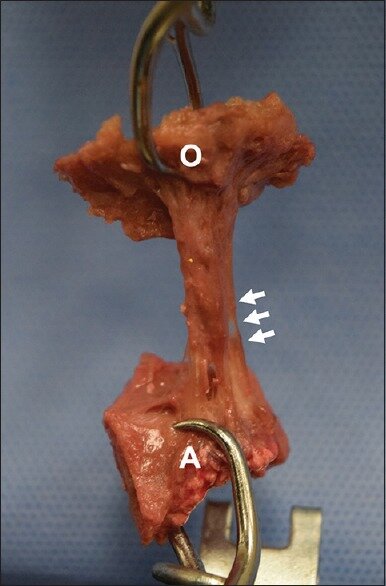
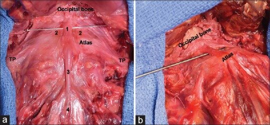
The superficial anterior atlanto-occipital ligament (SAAOL) is a narrowband located anterior to the anterior atlanto-occipital membrane. Nearly forgotten, it has not been well described in older anatomical textbooks and is missing in the current anatomical literature. As all of the binding structures of the craniocervical junction (CCJ) are important in maintaining stability, this study aims to clarify the anatomy and potential function of the SAAOL.

MATERIALS AND METHODS:

The CCJ from ten fresh-frozen cadavers was studied. These specimens were derived from three males and seven females, and the age at death ranged from 57 to 91 years (mean, 79.8 years). The length, width, and thickness of the SAAOL were measured. In five specimens, the force to failure was recorded.

RESULTS:

The SAAOL was found between the anterior tubercle of the atlas and the occiput and located as central thick fibers in front of the anterior atlanto-occipital membrane in 9 (90%) specimens. In one specimen, the vertical band to the occipital bone did not attach to the anterior tubercle of the atlas, but extended to the anterior aspect of the axis. The mean length, width, and thickness of the SAAOL were 19.8, 6.2, and 0.6 mm, respectively. The force to failure for the ligament was 38.8 N.

CONCLUSION:

The SAAOL was a constant structure of the anterior atlanto-occipital joint. This ligament seems to be a secondary stabilizer of the CCJ by limiting the extension of CCJ. Knowledge of this ligament may help in further understanding of craniocervical stability.

KEYWORDS:

Anatomy; cadaver; cervical vertebra; craniocervical joint; ligaments

Transclival Venous Circulation: Anatomic Study.

World Neurosurg. 2019 Jan;121:e136-e139. doi: 10.1016/j.wneu.2018.09.038. Epub 2018 Sep 18.

Altafulla JJRai RShrager SVoin VIwanaga JLitvack ZLoukas MTubbs RS.


Abstract

INTRODUCTION:

The clivus is a small, central area of the basal cranium with limited surgical access and high morbidity associated with pathologies of its surrounding structures. Therefore thorough knowledge and understanding of the anatomy in this region are crucial for the success of treatments and interpretation of imaging. As to our knowledge, there is no extant cadaveric examination of the transclival veins, so the present study was performed.

METHODS:

Fifteen lightly embalmed adult heads underwent blue latex injection of the left and right internal jugular veins. Special attention was given to the presence or absence of transclival vessels. When transclival veins were identified, their intracranial source, point of penetration of the clivus and anterior connections were documented.

RESULTS:

Ten (66.7%) specimens were found to have transclival veins. These connected the basilar venous plexus to the retropharyngeal venous plexus on all specimens. Eight of the 10 specimens had multiple transclival veins, and 2 had only 1 vessel. The majority of the transclival veins were found penetrating the clivus at its lower one third. However, 2 specimens also had transclival veins that pierced the clivus at its upper one third.

CONCLUSIONS:

An improved understanding of the skull base and its venous drainage can assist clinicians and surgeons in better understanding normal, pathologic, and variant anatomy in this region.

Copyright © 2018 Elsevier Inc. All rights reserved.

KEYWORDS:

Anatomy; Clival diploic veins; Clivus; Emissary veins; Transclival veins