The Diploic Veins: A Comprehensive Review with Clinical Applications.

Cureus. 2019 Apr 9;11(4):e4422. doi: 10.7759/cureus.4422.

Lachkar SDols MMIshak BIwanaga JTubbs RS.


Abstract

The diploic veins serve as an important connection between the extracranial and intracranial venous systems. They change in size during growth from adolescence to adulthood. The diploic space has been identified as an additional site of reabsorption of cerebrospinal fluid (CSF). Herein, the anatomy and physiology of the diploic veins are reviewed.

KEYWORDS:

arteriovenous fistula; cerebrospinal fluid; diploic space; diploic veins; diploic venous system

A Traumatic Dislocation of the Atlas from the Axis in a Patient with Atlantooccipital Assimilation.

Cureus. 2019 Apr 6;11(4):e4402. doi: 10.7759/cureus.4402.

Gnanadev RIshak BIwanaga JLoukas MTubbs RS.


Abstract

Atlantooccipital assimilation is defined as the complete or partial fusion of the caudal portion of the occiput with the cranial portion of the atlas. We report an extremely rare case of a traumatic dislocation of the atlas from the axis (C1 from C2), which lead to internal decapitation in a patient with atlantooccipital assimilation. This report reviews the background of atlantooccipital assimilations and their clinical relevance.

KEYWORDS:

assimilation; congenital; craniocervical; trauma

The Accessory Nerve: A Comprehensive Review of its Anatomy, Development, Variations, Landmarks and Clinical Considerations.

Anat Rec (Hoboken). 2019 Apr;302(4):620-629. doi: 10.1002/ar.23823. Epub 2018 Apr 23.

Johal JIwanaga JTubbs KLoukas MOskouian RJTubbs RS.


Abstract

INTRODUCTION:

The eleventh cranial nerve, the accessory nerve, has a complex and unique anatomy and has been the subject of much debate. Herein, we review the morphology, embryology, surgical anatomy, and clinical manifestations of the accessory nerve. Included in this review, we mention variant anatomy, molecular development, histology, and imaging of the accessory nerve.

CONCLUSIONS:

The accessory nerve continues to be a topic of much discussion regarding its exact function and in particular to its cranial roots. Recently, various surgical procedures have been devised that repurpose the accessory nerve (e.g., lengthening procedures, contralateral neurotization procedures). Currently, we continue to learn and have much to learn about this lower cranial nerve. Anat Rec, 302:620-629, 2019. © 2018 Wiley Periodicals, Inc.

© 2018 Wiley Periodicals, Inc.

KEYWORDS:

accessory nerve; cranial nerve; sternocleidomastoid; trapezius

Revisiting the genital and femoral branches of the genitofemoral nerve: Suggestion for a more accurate terminology.

Clin Anat. 2019 Apr;32(3):458-463. doi: 10.1002/ca.23327.

Iwanaga JSimonds ESchumacher MKikuta SWatanabe KTubbs RS.


Abstract

The genitofemoral nerve is a branch of the lumbar plexus originating from the ventral rami of the first and second lumbar spinal nerves. During routine dissections of this nerve, we have occasionally observed that the genital branch of the genitofemoral nerve gave rise to the femoral branch, and the femoral branch of the genitofemoral nerve gave rise to the genital branch. Therefore, this study aimed to investigate the aforementioned distributions of the genitofemoral nerve in a large number of cadaveric specimens. Twenty-four sides from fourteen fresh-frozen cadavers derived from nine males and five females were used in this study. For proximal branches of the genitofemoral nerve, that is, as they first arise from the genitofemoral nerve, the terms "medial branch" and "lateral branch" were used. For the final distribution, the terms "genital branch" and "femoral branch" were used. On eight sides (33.3%) with nine branches, one or two branch(s) from either the medial or lateral branch became coursed as the femoral or genital branches (five became femoral and four became genital branches). Our study revealed that the distribution of the genitofemoral nerve is more complicated than previously described. The "medial branch" and "lateral branch" that we have used in the present study for describing the proximal branches of the genitofemoral nerve are more practical terms to describe the genitofemoral nerve. Clin. Anat. 32:458-463, 2019.

© 2019 Wiley Periodicals, Inc.

KEYWORDS:

hernia; iatrogenic injury; inguinal canal; inguinal ligament; lumbar plexus; nerve block; neuralgia; pain

Human embryonic stem cells: Distinct molecular personalities and applications in regenerative medicine.

Clin Anat. 2019 Apr;32(3):354-360. doi: 10.1002/ca.23318. Epub 2019 Jan 7.

Dupont GYilmaz ELoukas MMacchi VDe Caro RTubbs RS.


Abstract

The field of stem cell biology is exciting because it provides researchers and clinicians with seemingly unlimited applications for treating many human diseases. Stem cells are a renewable source of pluripotent cells that can differentiate into nearly all human cell types. In this article we focus particularly on human embryonic stem (hES) cells, derived from the inner cell mass of the blastocyst and cultured for expansion while remaining undifferentiated, to explore their unique molecular personalities and clinical applications. The aim of this literature review is to reflect the interest in hES cells and to provide a resource for researchers and clinicians interested in the molecular characteristics of such cells. Clin. Anat. 32:354-360, 2019. © 2018 The Authors. Clinical Anatomy published by Wiley Periodicals, Inc. on behalf of American Association of Clinical Anatomists.

© 2018 The Authors. Clinical Anatomy published by Wiley Periodicals, Inc. on behalf of American Association of Clinical Anatomists.

KEYWORDS:

OCT4; disease; human embryonic stem cells; molecular; pluripotency; regeneration; stem cell biology; treatment

The anatomy of concussion and chronic traumatic encephalopathy: A comprehensive review.

Clin Anat. 2019 Apr;32(3):310-318. doi: 10.1002/ca.23313. Epub 2018 Dec 21.

Singla ALeineweber BMonteith SOskouian RJTubbs RS.


Abstract

Concussion is defined as a complex pathophysiological process that affects the brain as a result of traumatic biomechanical forces. Repeated unrecovered concussions can result in chronic brain injury syndrome which is referred to in the literature today as chronic traumatic encephalopathy." There is an exponential increase in public and political interest in this condition in the recent times resulting in a significant investment in research to improve the current understanding of the disease, ways to decrease its incidence and determine its prognosis. Broadly the research involves three main disciplines of medicine including neuropathology, neuroradiology and biological markers. Although progress has been made, to date there is no definite pathological, radiological or neurobiological marker which has shown consistent promise to make the diagnosis and prognosticate the disease. Possible reasons are multiple such as inconsistencies in the methods studies have used, different time periods in which the tests were conducted, the small numbers of subjects included in the studies, and inconsistencies in the definitions of concussion or mild traumatic brain injury. Herein, we present a comprehensive review of the current literature on this topic. Positron emission tomography scans with radioactive ligands such as T807 as an imaging biomarker, and neurofilament light and ubiquitin C-terminal hydrolase as serum biomarkers have shown some promise lately in diagnosing concussion and chronic traumatic encephalopathy and also determining their prognosis. Clin. Anat. 32:310-318, 2019.

© 2018 Wiley Periodicals, Inc.

KEYWORDS:

NFL; brain; football; guidelines; head; injury; return to play; sports; treatment

Modern operative nuances for the management of eloquent high-grade gliomas.

J Neurosurg Sci. 2019 Apr;63(2):135-161. doi: 10.23736/S0390-5616.18.04594-0. Epub 2018 Sep 25.

Mortazavi MMAhmadi Jazi GSadati MZakowicz KSheikh SKhalili KAdl FHTaqi MANguyen HSTubbs RS.


Abstract

INTRODUCTION:

Despite advancements in the treatment of high-grade gliomas (HGG), the rate of tumor recurrence is high and survival rate for the patient is low. Gross total resection has shown increased survival but the location of the tumor in the eloquent brain poses significant risk of morbidity. In this report, we focus on modern surgical nuances for resection of tumors located in the eloquent brain.

EVIDENCE ACQUISITION:

Research of the literature was conducted using the following search terms: surgical resection of gliomas, high-grade gliomas, and the role of vascular encasement - from 1986-2018. An institutional experience from the first author of this paper was also reviewed for selection of our illustrative cases.

EVIDENCE SYNTHESIS:

Gross total resection remains the mainstay of therapy for high-grade gliomas. The resection of the peritumoral FLAIR, when possible, has been associated with increased survival but also has the potential to cause increased morbidity. In the eloquent brain, the resection of the tumor itself is possible if attention is given to the interface of the tumor and brain, or if a safe pseudo-interface is created by the surgeon. Tumor-seeding to the ventricular system needs to be avoided. Devascularization, dissection away from the brain, and retractorless brain surgery are key to successful surgical outcomes. Management of the venous and arterial invasion/encasement are also outlined in this report. Technical aspects are discussed with corresponding videos.

CONCLUSIONS:

High-grade gliomas involving eloquent brain areas require a tailored treatment plan. While the medical treatment is undergoing quick evolution, gross total resection still remains one of the key milestones of treatment for improved survival. Surgical techniques play key role. We propose that encasement and/or the invasion of arteries and veins, should be considered equally as important as the eloquent brain when contemplating the resection of gliomas.

Guest UserComment
Neck movement during cervical transforaminal epidural injections and the position of the vertebral artery: an anatomical study.

Acta Radiol Open. 2019 Mar 12;8(3):2058460119834688. doi: 10.1177/2058460119834688. eCollection 2019 Mar.

Altafulla JYilmaz ELachkar SIwanaga JPeacock JLitvack ZTubbs RS.


Abstract

BACKGROUND:

Cervical transforaminal epidural steroid injections (CTFESIs) are sometimes performed in patients with cervical radiculopathy secondary to nerve-root compression. Neck movements for patient positioning may include rotation, flexion, and extension. As physicians performing such procedures do not move the neck for fear of injuring the vertebral artery, we performed fluoroscopy and cadaveric dissection to analyze any movement of the vertebral artery during head movement and its relation to the foramina in the setting of CTFESI.

PURPOSE:

To determine cervical rotational positioning for optimized vertebral artery location in the setting of cervical transforaminal epidural steroid injections.

MATERIAL AND METHODS:

Four sides from two Caucasian whole cadavers (all fresh-frozen) were used. Using a guide wire and digital subtraction fluoroscopy, we evaluated the vertebral artery mimicking a CTFESI, then we removed the transverse processes and evaluated the vertebral artery by direct observation.

RESULTS:

After performing such maneuvers, no displacement of the vertebral artery was seen throughout its course from the C6 to the C2 intervertebral foramina. To our knowledge, this is the first anatomical observation of its kind that evaluates the position of the vertebral artery inside the foramina during movement of the neck.

CONCLUSION:

Special caution should be given to the medial border of the intervertebral foramina when adjusting the target site and needle penetration for the injection. This is especially true for C6-C4 levels, whereas for the remaining upper vertebrae, the attention should be focused on the anterior aspect of the foramen. Since our study was centered on the vertebral artery, we do not discard the need for contrast injection and real-time digital subtraction fluoroscopy while performing the transforaminal epidural injection in order to prevent other vascular injuries.

KEYWORDS:

Epidural injection; cervicalgia; neck rotation; vertebral artery

Le Coup de Poignard Rachidien: A Historical Perspective.

Cureus. 2019 Mar 4;11(3):e4175. doi: 10.7759/cureus.4175.

Omar NBMiller JShoja MMHarrigan MRTubbs RS.


Abstract

A spinal subarachnoid hemorrhage (SAH) is uncommon. One of the earliest detailed analyses of a spinal SAH was in 1928 by the French physician Paul Michon, who coined the term "le coup de poignard rachidien" to describe the pathognomonic, intense spinal pain experienced by patients with spinal SAH, equating it to being stabbed by a dagger. Michon sub-classified spinal SAH into the upper and lower forms, pointing out that the stabbing spinal pain is more characteristic of SAH in the cervical and thoracic regions and especially in the interscapular region. Translation and subsequent analysis of Michon's original French paper published in La Presse Medicale in 1928 shed light on two cases in which patients presented with le coup de poignard rachidien and signs of spinal cord dysfunction but little, if any, intracranial symptoms. The patients both showed symptomatic relief following therapeutic lumbar puncture. Later, authors have questioned the notion that intense spinal or interscapular pain is mandatory in the diagnosis of spinal SAH and have additionally provided evidence contrary to Michon's assertion that intracranial symptoms, if any, occur later in the progression of spinal SAH and are largely insignificant.

KEYWORDS:

hemorrhage; michon; spinal; subarachnoid

Guest UserComment
An elongated dorsally curved xiphoid process.

Anat Cell Biol. 2019 Mar;52(1):102-104. doi: 10.5115/acb.2019.52.1.102. Epub 2019 Mar 29.

Lachkar SIwanaga JTubbs RS.


Abstract

The xiphoid process of the sternum lies in the epigastric region and functions to serve as an attachment point for vital muscles that aid in respiration. With the xiphoid process extending as the most inferior portion of the sternum, variable morphology is widely observed. During a routine dissection of a 44-year-old Caucasian male cadaver, we discovered a hook-shaped, elongated xiphoid process that protruded dorsally. Potential clinical significance can arise leading to misdiagnosis of the hook-shaped xiphoid process as an epigastric mass during imaging. Though various variations of xiphoid process have been well documented, knowledge of a hook-shaped xiphoid process orientated dorsally remains scarce. Herein, this case study provides clinicians, surgeons, and radiologists a rare anomaly of the xiphoid process in order to further the knowledge of morphological variations of the xiphoid to prevent misdiagnosis and surgical complications.

KEYWORDS:

Anatomic variation; Anatomy; Cadaver; Sternum; Xiphoid process

Report of an inferior rectal nerve variant arising from the S3 ventral ramus.

Anat Cell Biol. 2019 Mar;52(1):100-101. doi: 10.5115/acb.2019.52.1.100. Epub 2019 Mar 29.

Dupont GIwanaga JOskouian RJTubbs RS.


Abstract

In surgical approaches to the perineum in general and anal region specifically, considering the possible variations of the inferior rectal nerve is important for the surgeon. Normally, the inferior rectal nerve originates as a branch of the pudendal nerve. However, during routine dissection, a variant of the inferior rectal nerve was found where it arose directly from the third sacral nerve ventral ramus (S3). Many cases have described the inferior rectal nerve arising independently from the sacral plexus, most commonly from the fourth sacral nerve root (S4); however, few cases have reported the inferior rectal nerve arising from S3. Herein, we describe a variant of the inferior rectal nerve in which the nerve arises independently from the sacral plexus.

KEYWORDS:

Anal canal; Inferior rectal nerve variations; Perineum; Pudendal nerve block

Variant anatomy of the buccal nerve.

Anat Cell Biol. 2019 Mar;52(1):82-83. doi: 10.5115/acb.2019.52.1.82. Epub 2019 Mar 29.

Patel MIwanaga JKikuta SOskouian RJTubbs RS.


Abstract

Knowledge of the anatomy and variations of the nerves of the oral cavity is important to surgeons who operate this region. Herein, we report a rare case of a buccal nerve with two distinct roots. The anatomy of this case and its clinical applications is discussed.

KEYWORDS:

Anatomical variation; Anatomy; Buccal nerve; Buccal nerve block; Trigeminal nerve

Clinical anatomy of the maxillary sinus: application to sinus floor augmentation.

Anat Cell Biol. 2019 Mar;52(1):17-24. doi: 10.5115/acb.2019.52.1.17. Epub 2019 Mar 29.

Iwanaga JWilson CLachkar STomaszewski KAWalocha JATubbs RS.


Abstract

The anatomy of the maxillary sinus, especially its vascular anatomy, and its relationships with the teeth and alveolar processes have been well documented. The development of cone-beam computed tomography has resulted in dentists being more familiar with maxillary sinus floor augmentation procedures. This paper aims to revisit the classic anatomy of the maxillary sinus and review the newly published literature in order to help dentists diagnose in more detail and perform safer surgery of the maxillary sinus.

KEYWORDS:

Anatomy; Cadaver; Dental implants; Maxillary artery; Maxillary sinus; Sinus floor augmentation

Clinical anatomy of the nerve to the mylohyoid.

Anat Cell Biol. 2019 Mar;52(1):12-16. doi: 10.5115/acb.2019.52.1.12. Epub 2019 Mar 29.

Choi PIwanaga JDupont GOskouian RJTubbs RS.


Abstract

The nerve to the mylohyoid (NM) originates from the mandibular division of the trigeminal nerve. The NM provides motor control to the mylohyoid and the anterior belly of the digastric. Its sensory component, as a variation of this nerve, has scantly been described in the literature. We discuss the current clinical implications of the NM based on its anatomical variations of the with the hopes of benefiting patients who are undergoing invasive maxillofacial procedures.

KEYWORDS:

Anatomy; Inferior alveolar nerve; Mandibular nerve; Maxillofacial surgery; Nerve block; Variation.

Pediatric glossopharyngeal neuralgia: a comprehensive review.

Childs Nerv Syst. 2019 Mar;35(3):395-402. doi: 10.1007/s00381-018-3995-3. Epub 2018 Oct 25.

Shereen RGardner BAltafulla JSimonds EIwanaga JLitvack ZLoukas MShane Tubbs R.


Abstract

INTRODUCTION:

Debilitating facial pain can seriously affect an individual's daily living. Given that the pathophysiology behind neuropathic and myofascial pain is not fully understood, when chronic facial pain goes undiagnosed, it has been proposed that one of the two is the likely cause. Since their discovery, glossopharyngeal neuralgia (GN) and Eagle's syndrome have been considered mostly conditions afflicting the adult population. However, when pediatric patients present with symptoms resembling GN or Eagle's syndrome, physicians are less apt to include these as a differential diagnosis simply due to the low prevalence and incidence in the pediatric population.

MATERIALS AND METHODS:

A literature review was performed with the aim to better understand the history of reported cases and to provide a comprehensive report of the anatomical variations that lead to these two conditions as well as the way these variations dictated medical and surgical management. Articles were obtained through Google Scholar and PubMed. Search criteria included key phrases such as pediatric glossopharyngeal neuralgia and pediatric Eagle syndrome. These key phrases were searched independently. PubMed was searched primarily then cross-referenced articles were found via Google Scholar. Results from non-English articles were excluded.

RESULTS:

A total of 58 articles were reviewed. Most of the articles focused on adult glossopharyngeal neuralgia, and the majority was comprised of case reports. When searched via PubMed, a total of 16 articles and 2 articles returned for glossopharyngeal neuralgia and Eagle's syndrome, respectively. After criteria selection and cross-referencing, a total of seven articles were found with respect to pediatric glossopharyngeal neuralgia.

CONCLUSIONS:

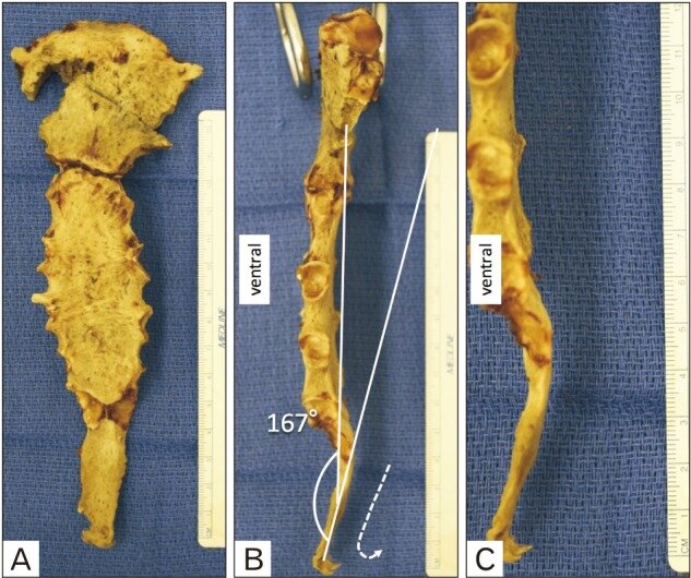
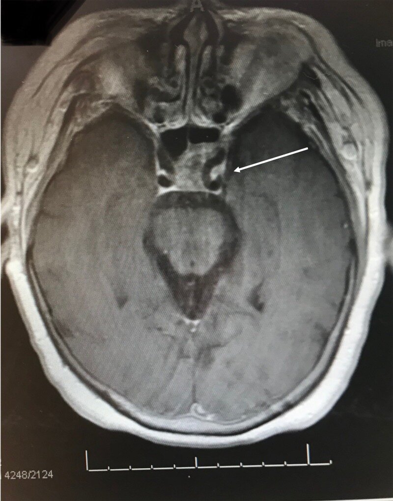
While they are rare conditions, there are multiple etiologies that lead to the debilitating symptoms of GN and Eagle's syndrome. The clinical anatomy proved notable as multiple causes of GN and Eagle's syndrome are due to variation in the anatomy of the neurovascular structures surrounding the glossopharyngeal nerve, an elongated styloid process, a calcified stylohyoid ligament as well as a calcified stylomandibular ligament. Due to the success of different treatment modalities, the treatment of choice is dependent on clinical judgment.

KEYWORDS:

Eagle’s syndrome; Gamma knife radiosurgery (GKRS); Glossopharyngeal neuralgia; Myofascial pain; Neuropathic; Trigeminal neuralgia

The majority of piriformis muscles are innervated by the superior gluteal nerve.

Clin Anat. 2019 Mar;32(2):282-286. doi: 10.1002/ca.23311. Epub 2018 Dec 21.

Iwanaga JEid SSimonds ESchumacher MLoukas MTubbs RS.


Abstract

The piriformis muscle is clinically implicated in pain disorders, posterior approaches for total hip arthroplasty, and iatrogenic injury to the muscle and the surrounding nerves. The piriformis muscle has been said to receive innervation from L5 to S3 ventral rami with most sources using S1 and S2 ventral rami as the most common innervation this muscle. However, descriptions of the nerve in the literature are vague. Therefore, the aim of this study was to clarify the anatomy of the nerve supply to the piriformis muscle. Twenty sides from ten fresh-frozen cadavers were studied. Specifically, via anterior dissection of the sacral plexus, branches to the piriformis were identified. Once identified, the nerves to the piriformis muscle were traced proximally to clarify their origin. Nerves supplying the piriformis muscle existed on all sides. On 80% of sides, the piriformis was innervated by two to three nerves. The origin of these nerves was from the superior gluteal nerve on 14 sides (70%), inferior gluteal nerve on one side (5%), L5 ventral ramus on one side (5%), S1 ventral ramus on 17 sides (85%), and S2 ventral ramus on 14 sides (70%), respectively. The most common nerve branches to the piriformis are from the superior gluteal nerve, and the ventral rami of S1 and S2. Based on our study, a single "nerve to piriformis" does not exist in the majority of specimens thus this term should be abandoned. Clin. Anat. 32:282-286, 2019.

© 2018 Wiley Periodicals, Inc.

KEYWORDS:

anatomy; cadaver; low back pain; piriformis syndrome; sacral plexus; sacroiliac joint; sciatica

Innervation of the anconeus epitrochlearis muscle: MRI and cadaveric studies.

Clin Anat. 2019 Mar;32(2):218-223. doi: 10.1002/ca.23285. Epub 2018 Oct 14.

Grewal SSCollin PIshak BIwanaga JAmrami KKRingler MDde Ruiter GCWSpinner RJTubbs RS.


Abstract

Ulnar neuropathy at the cubital tunnel is common. However, a rare form of ulnar neuropathy here is due to compression from an accessory muscle, the anconeus epitrochlearis. Reports in the literature regarding the details of this muscle's innervation are vague, so the aim of the present study was to characterize this anatomy more clearly. This was a combined review of magnetic resonance imaging (MRI) from patients with an anconeus epitrochlearis and ulnar neuropathy and cadaveric dissections to characterize the innervation of this variant muscle. A review of 11 patients and three reports of ulnar neuropathy and an anconeus epitrochlearis in the literature revealed no MRI changes consistent with acute denervation of this muscle. However, in two cases, there were signs of chronic denervation of the muscle. Dissection of five cadavers revealed that the nerve supply to the anconeus epitrochlearis originated proximal to the medial epicondyle, traveled parallel to the ulnar nerve, terminated on the deep aspect of this muscle, and had a mean length of 60 mm. This clinicoanatomical study provides evidence that the innervation of the anconeus epitrochlearis is proximal to the muscle and on its deep aspect. Clin. Anat. 32:218-223, 2019.

© 2018 Wiley Periodicals, Inc.

KEYWORDS:

anatomy; imaging; nerve compression; nervous; neuropathy; ulnar nerve

Ectopic Pituitary Adenoma Presenting as a Clival Mass.

Cureus. 2019 Feb 28;11(2):e4158. doi: 10.7759/cureus.4158.

Altafulla JJPrickett JTDupont GTubbs RSLitvack Z.


Abstract

Pituitary adenomas are well described in the literature and are frequently observed and treated in clinical practice by neurosurgeons. On the other hand, ectopic adenomas are a diagnostic enigma; a good understanding of anatomy and radiological characteristics is crucial for the successful management of such pathologies. In this paper, we describe the case of a 77-year-old woman who presented with a clival mass invading the left cavernous sinus; we also discuss the associated diagnostic techniques, approaches, imaging options, and characteristics.

KEYWORDS:

clival mass; histology; pituitary adenoma