An anatomical study of the lingual nerve in the lower third molar area.

Anat Cell Biol. 2019 Jun;52(2):140-142. doi: 10.5115/acb.2019.52.2.140. Epub 2019 Jun 30.

Kikuta SIwanaga JKusukawa JTubbs RS.


Abstract

The lingual nerve (LN) is a branch of the mandibular division of the trigeminal nerve, and its injury is one of the major complications during oral surgery. This study aims to investigate the anatomy of the LN in the lower third molar area. Twenty sides from ten fresh-frozen adult cadaveric Caucasian heads were examined to measure the diameter of the LN. The mean diameter of the LN was 2.20±0.37 mm (range, 1.61-2.95 mm). There were no statistically significant differences in the measurements between sexes, sides, or tooth status (dentulous or edentulous). Understanding the anatomical features of the LN is essential for performing any surgical procedure in the oral region.

KEYWORDS:

Anatomy; Fresh cadaver; Lingual nerve; Mandibular nerve; Wisdom tooth

Triangles of the neck: a review with clinical/surgical applications.

Anat Cell Biol. 2019 Jun;52(2):120-127. doi: 10.5115/acb.2019.52.2.120. Epub 2019 Jun 30.

Kikuta SIwanaga JKusukawa JTubbs RS.


Abstract

The neck is a geometric region that can be studied and operated using anatomical triangles. There are many triangles of the neck, which can be useful landmarks for the surgeon. A better understanding of these triangles make surgery more efficient and avoid intraoperative complications. Herein, we provide a comprehensive review of the triangles of the neck and their clinical and surgical applications.

KEYWORDS:

Anatomy; Landmarks; Neck; Surgery; Triangle

Ossification of the mamillo-accessory ligament: a review of the literature and clinical considerations.

Anat Cell Biol. 2019 Jun;52(2):115-119. doi: 10.5115/acb.2019.52.2.115. Epub 2019 Jun 30.

Dupont GYilmaz EIwanaga JOskouian RJTubbs RS.


Abstract

Ossification of the mamillo-accessory ligament (MAL) is a misunderstood phenomenon; however, many have posited that it can result in nerve entrapment of the medial branch of the dorsal ramus causing zygapophyseal joint related low back pain. The MAL has been studied anatomically by few, yet the data indicate possible associations between ossification of this ligament and spondylosis. It has been proposed that mechanical stress upon the lumbar spine may also lead to progressive ossification of the MAL into a bony foramen.

KEYWORDS:

Anatomy; Low back pain; Lumbar; Mamillo-accessory ligament; Medial branch block; Ossification

Correction to: Untreated incomplete isolated cleft palate: cadaveric findings.

Anat Sci Int. 2019 Jun;94(3):274. doi: 10.1007/s12565-019-00484-4.

Kikuta SIwanaga JKusukawa JOskouian RJTubbs RS.


Abstract

In the original publication, the given name and family name of the last author was incorrectly published. The correct given name and family name should be "R. Shane" and "Tubbs".

Erratum for

Guest UserComment
Nerve to mylohyoid branched from the lingual nerve: previously undescribed case.

Anat Sci Int. 2019 Jun;94(3):266-268. doi: 10.1007/s12565-019-00476-4. Epub 2019 Feb 1.

Iwanaga JKikuta SOskouian RJTubbs RS.

Abstract

The lingual nerve is a branch of the mandibular division of the trigeminal nerve. It descends medial and anterior to the inferior alveolar nerve through the pterygomandibular space, runs by the lingual plate and lingual crest at the lower third molar closely, and supplies sensory fibers to the anterior two-thirds of the tongue. Therefore, injury of this nerve is occasionally induced by wisdom tooth extraction and could lead to paralysis of the tongue. The inferior alveolar nerve gives rise to the nerve to mylohyoid just before entering the mandibular foramen, which supplies the mylohyoid and anterior belly of the digastric muscle. We present an extremely rare anatomical variation where the nerve to mylohyoid arose from the lingual nerve near the submandibular duct during routine oral dissection.

KEYWORDS:

Anatomy; Cadaver; Lingual nerve; Mandibular nerve; Wisdom tooth

A comprehensive review of the sinuvertebral nerve with clinical applications.

Anat Cell Biol. 2019 Jun;52(2):128-133. doi: 10.5115/acb.2019.52.2.128. Epub 2019 Jun 30.

Shayota BWong TLFru DDavid GIwanaga JLoukas MTubbs RS.


Abstract

The anatomy and clinical significance of the sinuvertebral nerve is a topic of considerable interest among anatomists and clinicians, particularly its role in discogenic pain. It has required decades of research to appreciate its role, but not until recently could these studies be compiled to establish a more complete description of its clinical significance. The sinuvertebral nerve is a recurrent nerve that originates from the ventral ramus, re-entering the spinal canal via the intervertebral foramina to innervate multiple meningeal and non-meningeal structures. Its complex anatomy and relationship to discogenic pain have warranted great interest among clinical anatomists owing to its sympathetic contribution to the lumbar spine. Knowledge of the nerve has been used to design a variety of diagnostic and treatment procedures for chronic discogenic pain. This paper reviews the anatomy and clinical aspects of the sinuvertebral nerve.

KEYWORDS:

Anatomy; Innervation; Meninges; Pain; Spine

Avoiding the Esophageal Branches of the Recurrent Laryngeal Nerve During Retractor Placement: Precluding Postoperative Dysphagia During Anterior Approaches to the Cervical Spine.

Global Spine J. 2019 Jun;9(4):383-387. doi: 10.1177/2192568218810198. Epub 2019 Feb 11.

Fisahn CYilmaz EIwanaga JSchmidt CBenca EChapman JROskouian RJTubbs RS.


Abstract

STUDY DESIGN:

Anatomical cadaver study.

OBJECTIVES:

Postoperative dysphagia is a significant complication following anterior approaches to the cervical spine and the etiology of this complication is poorly understood. Herein, we studied the esophageal branches of the recurrent laryngeal nerves to improve understanding of their anatomy and potential involvement in dysphagia.

METHODS:

Ten fresh frozen cadaveric human specimens were dissected (20 sides). All specimens were adults with no evidence of prior surgery of the anterior neck. The recurrent laryngeal nerves were identified under a surgical microscope and observations and measurements of their esophageal branches made.

RESULTS:

For each recurrent laryngeal nerve, 5-7 (mean 6.2) esophageal branches were identified. These branches ranged from 0.8 to 2.1 cm (mean 1.5 cm) in length and 0.5 to 2 mm (mean 1 mm) in diameter. They arose from the recurrent laryngeal nerves between vertebral levels T1 and C6. They all traveled to the anterior aspect of the esophagus. No statistical differences were seen between left and right sides or between sexes.

CONCLUSION:

The esophageal branches of the recurrent laryngeal nerve have been poorly described and could contribute to complications such as swallowing dysfunction following anterior cervical discectomy and fusion procedures. Therefore, a better understanding of their anatomy is important for spine surgeons. Our study revealed that these branches are always present on both sides and the anterior surface of the esophagus should be avoided while retracting it in order to minimize the risk of postoperative dysphagia.

KEYWORDS:

anatomy; anterior cervical discectomy; complications; dysphagia; neck; surgery; swallowing

Iatrogenic Bowel Injury Following Minimally Invasive Lateral Approach to the Lumbar Spine: A Retrospective Analysis of 3 Cases.

Global Spine J. 2019 Jun;9(4):375-382. doi: 10.1177/2192568218800045. Epub 2018 Nov 20.

Rustagi TYilmaz EAlonso FSchmidt COskouian RTubbs RSChapman JRHopkins SSchildhauer TAFisahn C.


Abstract

STUDY DESIGN:

Retrospective cohort study.

OBJECTIVE:

Anterior approaches are often used during lumbar interbody fusion procedures. Visceral injuries (bowel injuries) are rare but represent a primary risk during anterior approaches to the lumbar spine. Left untreated, these injuries can result in significant complications. The aim of this study was to investigate the presentation and management of bowel injury cases following anterior approaches to the lumbar spine to raise the surgeon's awareness of this rare complication.

METHODS:

All direct anterior, oblique anterior, and transpsoas lumbar interbody fusion surgeries performed at our institution between 2012 and 2016 were analyzed retrospectively. Charts were screened for cases requiring return to the operating room owing to a suspected bowel injury and details of the case were extracted for illustrative purposes.

RESULTS:

A total of 775 anterior lumbar surgeries were conducted at a single tertiary care institution between July 2012 and June 2017. A total of 590 transpsoas lumbar interbody fusion (TPIF) surgeries were performed. Four patients, each having undergone TPIF, were suspected of bowel injury and underwent an exploratory laparotomy. At surgery, 3 patients were confirmed to have a bowel injury, giving a procedure-specific incidence of 0.51% and overall incidence of 0.39%. Among the 3 confirmed bowel injury cases, average delay between surgery and visceral injury diagnosis was 4.7 days (range 3-7 days).

CONCLUSIONS:

We noted abdominal pain, distention, and fever as the most common findings in the setting of a visceral injury. A high index of suspicion and computed tomography imaging remain critical for identifying postoperative bowel injuries.

KEYWORDS:

bowel injury; complication; lateral approach; peritonitis; spine surgery

Buccal Nerve Dissection Via an Intraoral Approach: Correcting an Error Regarding Buccal Nerve Blockade.

J Oral Maxillofac Surg. 2019 Jun;77(6):1154.e1-1154.e4. doi: 10.1016/j.joms.2019.01.038. Epub 2019 Feb 5.

Iwanaga JTubbs RS.


Abstract

PURPOSE:

The buccal nerve (BN) supplies sensation to the skin over the anterior part of the buccinator, buccal mucosa, and buccal gingivae corresponding to the second and third molar teeth. Some dental anatomy books depict the main trunk of the BN coursing over the buccal shelf of the mandible, which leads to a serious misunderstanding of this anatomy. Therefore, this study aimed to show the course of the BN on the retromolar area and establish new evidence-based anatomy for appropriate BN blockade.

MATERIALS AND METHODS:

Twenty sides from 10 fresh-frozen Caucasian cadaveric heads were used in this study. The closest distance from the BN to the injection site of the BN blockade (Dis A) was measured.

RESULTS:

The mean Dis A was 12.69 ± 2.95 mm. On 16 of 20 sides (80%), Dis A was longer than 10 mm. In most specimens, the injection site of the BN blockade was too far from the course of the main trunk of the BN and the injection site should be changed to 10 mm superolateral to the injection site based on the present results and those of other previous studies.

CONCLUSIONS:

Although the distribution of the BN is variable, in most specimens, the current injection site for BN blockade was too far from the course of the BN.

Copyright © 2019 American Association of Oral and Maxillofacial Surgeons. Published by Elsevier Inc. All rights reserved.

Guest UserComment
Thoracolumbar Injury Classification and Severity Score in Children: A Validity Study.

Neurosurgery. 2019 Jun 1;84(6):E362-E367. doi: 10.1093/neuros/nyy408.

Dawkins RLMiller JHMenacho STRamadan OILysek MCKuhn ENTubbs RSWalker MLWalters BCAgee BSRozzelle CJ


Abstract

BACKGROUND:

The Thoracolumbar Injury Classification and Severity Score (TLICS) has been shown to be a valid tool for assessing the need for surgical intervention in adult patients. There is limited insight into its usefulness in children.

OBJECTIVE:

To assess the validity of the TLICS system in pediatric patients.

METHODS:

The medical records for pediatric patients with acute, traumatic thoracolumbar fractures at two Level 1 trauma centers were reviewed retrospectively. A TLICS score was calculated for each patient using computed tomography and magnetic resonance images, along with the neurological examination recorded in the patient's medical record. TLICS scores were compared with the type of treatment received. Receiver operating characteristic (ROC) curve analysis was employed to quantify the validity of the TLICS scoring system.

RESULTS:

TLICS calculations were completed for 165 patients. The mean TLICS score was 2.9 (standard deviation ± 2.7). Surgery was the treatment of choice for 23% of patients. There was statistically significant agreement between the TLICS suggested treatment and the actual treatment received (P < 0.001). The ROC curve calculated using multivariate logistic regression analysis of the TLICS system's parameters as a tool for predicting treatment demonstrated excellent discriminative ability, with an area under the ROC curve of 0.96, which was also statistically significant (P < 0.001).

CONCLUSION:

The TLICS system demonstrates good validity for selecting appropriate thoracolumbar fracture treatment in pediatric patients.

Copyright © 2018 by the Congress of Neurological Surgeons.

KEYWORDS:

Children; Fracture; Score; Thoracolumbar; Validity

The condylar canal and emissary vein-a comprehensive and pictorial review of its anatomy and variation.

Childs Nerv Syst. 2019 May;35(5):747-751. doi: 10.1007/s00381-019-04120-4. Epub 2019 Mar 21.

Lachkar SKikuta SIwanaga JTubbs RS.


Abstract

The condylar canal and its associated emissary vein serve as vital landmarks during surgical interventions involving skull base surgery. The condylar canal serves to function as a bridge of communication from the intracranial to extracranial space. Variations of the condylar canal are extremely prevalent and can present as either bilateral, unilateral, or completely absent. Anatomical variations of the condylar canal pose as a potential risk to surgeons and radiologist during diagnosis as it could be misinterpreted for a glomus jugular tumor and require surgical intervention when one is not needed. Few literature reviews have articulated the condylar canal and its associated emissary vein through extensive imaging. This present paper aims to further the knowledge of anatomical variations and surgical anatomy involving the condylar canal through high-quality computed tomography (CT) images with cadaveric and dry bone specimens that have been injected with latex to highlight emissary veins arising from the condylar canal.

KEYWORDS:

Anatomical variation; Anatomy; Cadaver; Emissary vein; Posterior condylar canal; Skull

Jaw pain and myocardial ischemia: A review of potential neuroanatomical pathways.

Clin Anat. 2019 May;32(4):476-479. doi: 10.1002/ca.23367. Epub 2019 Mar 22.

Kikuta SDalip DLoukas MIwanaga JTubbs RS.


Abstract

Cardiac pain is usually manifested as a crushing, squeezing, or sensation of pressure in the center of the chest. The pain can be referred to the left shoulder, neck, jaw, and epigastric region as well as the temporomandibular region, paranasal sinuses, and head in general. Although not well understood, during myocardial ischemia, the process of cardiac referred pain to craniofacial structures can be explained by the convergence of visceral and somatic relays at the trigeminal nucleus in the brain stem. The goal of this article is to review the possible pathways for referred jaw pain due to myocardial ischemia. Clin. Anat. 32:476-479, 2019.

© 2019 Wiley Periodicals, Inc.

KEYWORDS:

cardiac pain; convergence; myocardial ischemia; referred pain

Breathing Life into the Cadaver: Introducing Air Dissection As a New Teaching and Dissecting Method for the Venous System.

Clin Anat. 2019 May;32(4):566-572. doi: 10.1002/ca.23354. Epub 2019 Mar 7.

Iwanaga JKikuta STubbs RS.


Abstract

Air insufflation has been used for various surgical procedures such as during laparoscopy. We hypothesized that the use of pressurized air might enable cadaveric dissection to differentiate smaller veins better than traditional dissection techniques. In three fresh-frozen cadavers, the inferior vena cava (IVC) and right or left femoral veins were exposed just distal to the inguinal ligament and a needle placed into one of them. Pressurized air was then placed into the cannulated femoral vein using an air compressor. In all specimens, the IVC and most of its tributaries, both left and right sides, were clearly insufflated. When the IVC was traced superiorly by resecting the diaphragm through the caval foramen, the right atrium and ventricle were also found to be dilated. Additionally, venous variants that would have not been obvious without dilatation of the IVC were identified. Air dissection of the venous system in fresh-frozen cadavers aids in anatomical dissection. Such a model might also serve as a surgical training model and teaching tool as it better mimics life-like anatomy and physiology. We term this technique "cadaveric air dissection." Clin. Anat. 32:566-572, 2019.

© 2019 Wiley Periodicals, Inc.

KEYWORDS:

air dissection; anatomic variations; anatomy; cadaver; dissection; vein; venous system

A new variant ligament of the atlantooccipital joint: the lateral oblique atlantooccipital ligament.

Eur Spine J. 2019 May;28(5):1188-1191. doi: 10.1007/s00586-019-05919-0. Epub 2019 Feb 19.

Kikuta SIwanaga JOskouian RJTubbs RS.


Abstract

PURPOSE:

During routine dissection of the anterior craniocervical junction (CCJ), a variant ligament just anterior to the articular capsule of the atlantooccipital joint was observed. To our knowledge, no literature has previously described this ligament. Therefore, the aim of this study was to clarify the anatomy, incidence, and biomechanics of this undescribed structure of the anterior atlantooccipital joint.

METHODS:

Twenty-six sides from 13 fresh-frozen adult cadavers were used for this study and the morphology of the variant ligament examined. When present, its length, width, thickness, and the angle from the midline of the CCJ were measured.

RESULTS:

The variant ligament identified, when present, is distinct and located anterior to the atlantooccipital joint capsule traveling between the occipital bone and the transverse process of the atlas. The ligament was found on 12 of 26 sides (46.2%). The mean length of the ligament was 32.0 ± 5.5 mm. The ligament became taut with contralateral lateral flexion and the ipsilateral rotation of the atlantooccipital joint.

CONCLUSIONS:

We propose that this ligament may be termed the lateral oblique atlantooccipital ligament. To date, this structure has not been described in any textbooks or reports in the extant medical literature. Although its function is not clear, based on its course and connections, it might function as a secondary stabilizer of the atlantooccipital joint. As the stability of the craniocervical junction is of paramount importance, knowledge of normal and variant anatomical structures in this region is important for the surgeon treating patients with pathology of this region. These slides can be retrieved under Electronic Supplementary Material.

KEYWORDS:

Anatomy; Cadaver; Cervical vertebra; Craniocervical joint; Ligaments; Skull base

Anatomic Study of the Superior Cluneal Nerve and Its Related Groove on the Iliac Crest.

World Neurosurg. 2019 May;125:e925-e928. doi: 10.1016/j.wneu.2019.01.210. Epub 2019 Feb 11.

Iwanaga JSimonds ESchumacher MYilmaz EAltafulla JTubbs RS.


Abstract

OBJECTIVE:

Superior cluneal nerve entrapment neuropathy is one cause of low back pain often referred to as "pseudo sciatica." Studies have found that the superior cluneal nerve can arise variably from T11 to L5. The osteofibrous tunnels formed by a groove on the iliac crest might compress the superior cluneal nerve. Therefore, the purpose of this study was to investigate the origin of the superior cluneal nerve and its course through such bony grooves.

METHODS:

Twenty sides from 10 fresh frozen Caucasian cadavers were used in this study. Once both the superior cluneal nerve and its groove were identified, the distance from the groove to the posterior superior iliac spine and midline was measured.

RESULTS:

A total of 12 grooves were identified in 11 of 20 sides (55%). On 10 sides, the nerve running on the groove was the medial branch of the superior cluneal nerve. The mean distance from the bony groove to the posterior superior iliac spine and midline was 45.2 ± 11.2 mm and 65.3 ± 8.2 mm, respectively.

CONCLUSIONS:

These results could help identify such bony grooves and better understand low back pain and its related anatomy.

Copyright © 2019 Elsevier Inc. All rights reserved.

KEYWORDS:

Anatomy; Low back pain; Lumbar vertebrae; Nerve compression syndrome; Pseudo sciatica

The Great Auricular Nerve: Anatomical Study with Application to Nerve Grafting Procedures.

World Neurosurg. 2019 May;125:e403-e407. doi: 10.1016/j.wneu.2019.01.087. Epub 2019 Jan 28..

Altafulla JIwanaga JLachkar SPrickett JDupont GYilmaz EIshak BLitvack ZTubbs RS.


Abstract

BACKGROUND:

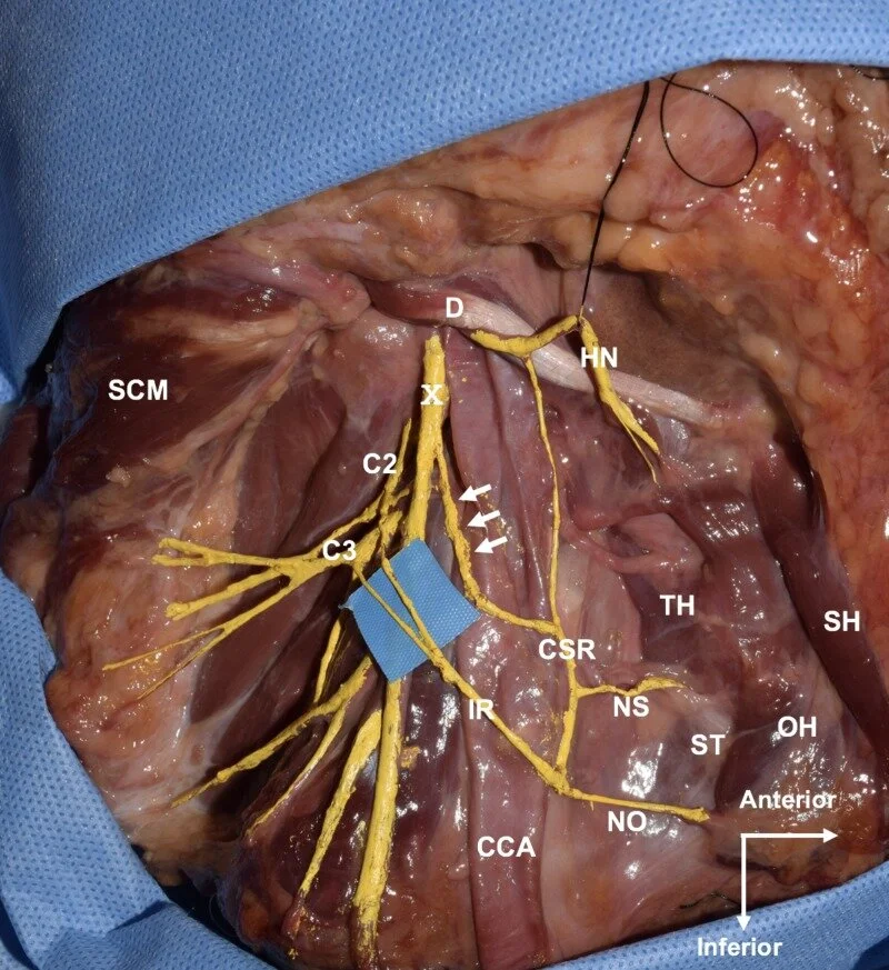
When it comes to autogenous nerve grafting, the sural and great auricular nerve (GAN) are the 2 nerves predominately used for trigeminal and facial nerve repair. Arising from the second and third cervical ventral rami, the GAN emerges from the posterior border of the sternocleidomastoid coursing superiorly and anteriorly toward the ear.

METHODS:

Eleven sides from 5 Caucasian and 1 Asian cadaveric heads (all fresh-frozen) were used. One man and 5 women were used with an age at death ranging from 57 to 91 years, with a mean of 80.3 years. Measurements were made from the inferior border of the ear to the GAN, the GAN to the external jugular vein, and the inferior border of the mastoid process to the GAN; the proximal, medial, and distal diameters of the GAN and the length of the GAN that was obtained from this exposure were also measured.

RESULTS:

The mean distance from the inferior border of the mastoid process to the GAN, inferior border of the ear to the GAN, and GAN to the external jugular vein was 27.71, 31.03, and 13.28 mm, respectively. The mean length of the GAN was 74.86 mm. The mean diameter of its distal, middle, and proximal portions was 1.51, 1.38, and 1.58 mm, respectively.

CONCLUSIONS:

The GAN is an excellent option for use in nerve grafting for repair of, for example, facial dysfunction. In this study, we review our measurements, techniques for identification, and dissecting techniques for the GAN. The proximity to the operative area and minimal complications associated with GAN grafting might contribute to improved patient satisfaction and better outcomes regarding functional restoration.

Copyright © 2019 Elsevier Inc. All rights reserved.

KEYWORDS:

External jugular vein; Great auricular nerve; Nerve graft; Sternocleidomastoid

Absence of the Right Common Iliac Vein with the Right Internal Iliac Vein Arising from the Left Common Iliac Vein: Case Report.

Cureus. 2019 Apr 30;11(4):e4575. doi: 10.7759/cureus.4575.

Mehta KIwanaga JTubbs RS.


Abstract

The common iliac vein arises from the internal and external iliac veins and drains into the inferior vena cava, bilaterally. Historically, many anatomical variants of the common iliac vein have been studied. To our knowledge, we discuss a unique specimen, which presents with an absent right common iliac vein and a right internal iliac vein draining into the contralateral common iliac vein. It is important that we understand the anatomic variations of the pelvic venous system to prevent iatrogenic injury to patients or misdiagnosis.

KEYWORDS:

anatomy; cadaver; common iliac vein; variation

An Unusual Superior Root of the Ansa Cervicalis.

Cureus. 2019 Apr 27;11(4):e4558. doi: 10.7759/cureus.4558.

Kikuta SIwanaga JKusukawa JTubbs RS.


Abstract

The ansa cervicalis is located around the carotid sheath and forms a neural loop, which consists of superior and inferior roots. It innervates the infrahyoid muscles. Anatomical variations of the superior root of the ansa cervicalis are uncommon. Herein, we present an extremely rare case of the superior root of the ansa cervicalis arising both from the hypoglossal and vagus nerves.

KEYWORDS:

anatomy; ansa cervicalis; neck surgery; vagus nerve; variation

Unusual Origin of the Anterior Scrotal Nerve: A Case Report.

Cureus. 2019 Apr 27;11(4):e4557. doi: 10.7759/cureus.4557.

Mehta KIwanaga JTubbs RS.


Abstract

The anterior scrotal nerve is a cutaneous nerve that branches from the ilioinguinal nerve after it leaves the superficial inguinal ring. However, we identified a cadaveric specimen with an anterior scrotal nerve arising from both the femoral and ilioinguinal nerves. This anatomic variation should be considered with anesthetic blockade of this region or during surgical procedures nearby.

KEYWORDS:

anatomy; cadaver; femoral nerve; ilioinguinal nerve; scrotum