Division of Sacrospinous and Sacrotuberous Ligaments Expands Access Through Greater Sciatic Foramen: Anatomic Study with Application to Resection of Greater Sciatic Foramen Tumors.

World Neurosurg. 2019 Aug;128:e970-e974. doi: 10.1016/j.wneu.2019.05.045. Epub 2019 May 14.

Iwanaga JPuffer RCWatanabe KSpinner RJTubbs RS.


Abstract

OBJECTIVE:

Tumors of the greater sciatic foramen remain difficult to treat. They often have both intrapelvic and extrapelvic components that may limit visualization and make safe resection of the tumor difficult. Therefore the goal of the present anatomic study was to quantitate how much additional surgical working space could be gained by transection of the sacrospinous and sacrotuberous ligaments.

METHODS:

Sixteen sides from 9 fresh-frozen Caucasian cadaveric torsos underwent transgluteal dissection and exposure of the greater sciatic foramen and associated liagments. With the piriformis in place, the vertical and horizontal diameters of the greater sciatic foramen were measured. Next, the sacrotuberous and sacrospinous ligaments were cut at their ischial attachments. The vertical diameter of the now confluent greater and lesser sciatic foramina (V2) was measured.

RESULTS:

The mean vertical diameter of the greater sciatic foramen (V1) was 54.8 ± 9.7 mm. The horizontal diameter of the greater sciatic foramen had a mean of 44.3 ± 6.1 mm with a range of 30-52 mm. After transection of the sacrotuberous and sacrospinous ligaments, the vertical distance of the greater and lesser sciatic foramina (V2) had a mean of 74.8 ± 6.8 mm with a range of 60.1-90 mm. The mean ratio of V2 to V1 was 1.40.

CONCLUSIONS:

The vertical length of the greater sciatic foramen increased, on average, 40% after resection of the sacrotuberous and sacrospinous ligaments. The results of this study support an alternative technique for resecting large intrapelvic tumors via a transgluteal approach.

Copyright © 2019 Elsevier Inc. All rights reserved.

KEYWORDS:

Dumbbell tumor; Greater sciatic foramen; Sacrospinous ligament; Sacrotuberous ligament; Sciatic nerve

Iliolumbar Vein: Anatomy and Surgical Importance During Lateral Transpsoas and Oblique Approaches to Lumbar Spine.

World Neurosurg. 2019 Aug;128:e768-e772. doi: 10.1016/j.wneu.2019.04.252. Epub 2019 May 9.

Davis MJenkins SBordes SIwanaga JLoukas MUribe JHynes RATubbs RS.


Abstract

OBJECTIVE:

Safe surgical approaches to the anterolateral lumbar spine require a good working knowledge of the anatomy and anatomic variations of this region. As the iliolumbar vein is in the vicinity of both oblique and lateral transpsoas approaches to the lower lumbar spine, the following study was performed to better elucidate its anatomy, variations, and position during such surgical procedures.

METHODS:

Fifteen (30 sides) fresh frozen adult cadavers underwent dissection of the iliolumbar vein (ILV). The origin, course, variants, relations, and morphometrics of each vein were documented. Fluoroscopy of the vessels was performed. Lastly, anterior oblique and lateral transpsoas approaches to the lumbar spine were carried out in order to evaluate for potential ILV injury.

RESULTS:

An ILV was found on all but 2 sides (93.3%). It arose as a common trunk from the common iliac vein on 14 sides. Left ILVs tended to have a more distal origin than right ILVs. ILVs had a mean length of 3.7 cm and a mean width of 0.9 cm and were significantly larger on right versus left sides (P < 0.05). Left-sided ILVs tended to have more branches than right-sided veins. The majority of vertical branches of the ILV traveled anterior to the ventral rami of the lumbar spinal nerves, most commonly L4. The ILV and, in particular, its vertical branches coursed next to the L4 and L5 vertebrae.

CONCLUSIONS:

The ILV should be considered during both oblique and lateral transpsoas approaches to the lumbar spine.

Copyright © 2019 Elsevier Inc. All rights reserved.

KEYWORDS:

Ascending lumbar vein; Iliolumbar vein; Lateral approach to spinal surgery; Vascular injury

Anatomic Study of Superior Cluneal Nerves: Revisiting the Contribution of Lumbar Spinal Nerves.

World Neurosurg. 2019 Aug;128:e12-e15. doi: 10.1016/j.wneu.2019.02.159. Epub 2019 Mar 9.

Iwanaga JSimonds ESchumacher MOskouian RJTubbs RS.


Abstract

OBJECTIVE:

Superior cluneal nerve (SCN) entrapment neuropathy can result in low back pain and thus be confused with other pathologies (e.g., lumbar disk disease). Therefore we performed cadaveric dissection of the SCN to better understand its anatomy and segmental origin.

METHODS:

Twenty sides from 10 Caucasian fresh frozen cadavers (6 females and 4 males) were used in this study. The diameter of the SCN, distance between the exit point of the SCN from the thoracolumbar fascia and midline, and distance between the exit point of the SCN from the thoracolumbar fascia and the posterior superior iliac spine to the medial and lateral SCN were measured. The segmental origins of the SCNs were verified.

RESULTS:

Seventy-five percent of the dorsal rami of L1, 90% of L2, 95% of L3, 45% of L4, and 10% of L5 contributed to the SCN. The SCN was formed by 3 vertebral levels in 55% and by 4 vertebral levels in 30%. Three SCNs pierced the thoracolumbar fascia in 45%.

CONCLUSIONS:

The origin of the SCN, which has been described in the textbook and literature for a long time, should be reconsidered on the basis of our study results.

Copyright © 2019 Elsevier Inc. All rights reserved.

KEYWORDS:

Anatomy; Low back pain; Lumbar vertebrae; Nerve compression syndrome; Pseudosciatica

Operative Intervention for Lumbar Foraminal Gunshot Wounds: Case Report and Review of the Literature.

Cureus. 2019 Jul 29;11(7):e5269. doi: 10.7759/cureus.5269.

Brash AHalalmeh DRRajah GLoya JMoisi M.


Abstract

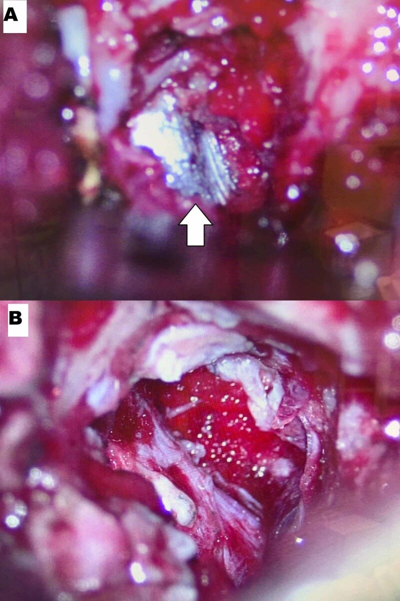
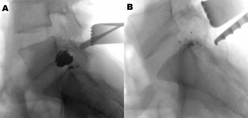
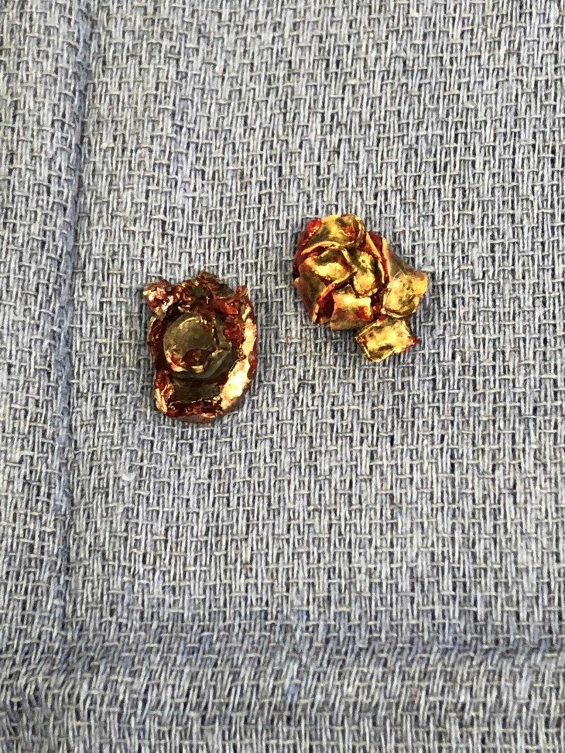
Gunshot wounds represent the second most frequent cause of spinal cord injury after vehicular trauma. The thoracic region is most commonly involved, followed by the thoracolumbar spine. Numerous studies have demonstrated that improvement of neurological recovery, especially after decompression surgery, is likely to be seen in lumbosacral spine, but not in the thoracic or cervical spine. Herein, we present a case of a gunshot wound causing lumbar 5th nerve root compression with neurological deficits that improved remarkably after urgent decompression surgery. This signifies a potential neurological benefit to prompt surgical intervention in lumbar gunshot wounds with radiographic evidence of neural compression. A relevant review of the literature was performed along with discussion, the clinical history, and radiological findings.

Copyright © 2019, Brash et al.

KEYWORDS:

bullet; gunshot wound; lumbar spine; treatment

Loss of consciousness at onset of aneurysmal subarachnoid hemorrhage in good-grade patients.

Neurosurg Rev. 2019 Jul 22. doi: 10.1007/s10143-019-01142-z. [Epub ahead of print]

Hendrix PForeman PMSenger SBurkhardt BWHarrigan MRFisher WS 3rdVyas NALipsky RHWalters BCTubbs RSShoja MMGriessenauer CJ.


Abstract

Loss of consciousness (LOC) at presentation with aneurysmal subarachnoid hemorrhage (aSAH) has been associated with early brain injury and poor functional outcome. The impact of LOC on the clinical course after aSAH deserves further exploration. A retrospective analysis of 149 aSAH patients who were prospectively enrolled in the Cerebral Aneurysm Renin Angiotensin Study (CARAS) between 2012 and 2015 was performed. The impact of LOC was analyzed with emphasis on patients presenting in excellent or good neurological condition (Hunt and Hess 1 and 2). A total of 50/149 aSAH patients (33.6%) experienced LOC at presentation. Loss of consciousness was associated with severity of neurological condition upon admission (Hunt and Hess, World Federation of Neurosurgical Societies (WFNS), Glasgow Coma Scale (GCS) grade), hemorrhage burden on initial head CT (Fisher CT grade), acute hydrocephalus, cardiac instability, and nosocomial infection. Of Hunt and Hess grade 1 and 2 patients, 21/84 (25.0%) suffered LOC at presentation. Cardiac instability and nosocomial infection were significantly more frequent in these patients. In multivariable analysis, LOC was the predominant predictor of cardiac instability and nosocomial infection. Loss of consciousness at presentation with aSAH is associated with an increased rate of complications, even in good-grade patients. The presence of LOC may identify good-grade patients at risk for complications such as cardiac instability and nosocomial infection.

KEYWORDS:

Aneurysm; Loss of consciousness; Subarachnoid hemorrhage

Disaster Scenarios in Spine Surgery: A Survey Analysis.

Spine (Phila Pa 1976). 2019 Jul 15;44(14):1018-1024. doi: 10.1097/BRS.0000000000003040.

Abdul-Jabbar AYilmaz EFisahn CDrazin DBlecher RUppal MPhernetton BLAltafulla JOskouian RJChapman JR.


Abstract

STUDY DESIGN:

Survey analysis among spine surgeons.

OBJECTIVE:

To identify current consensus and discrepancies in managing adverse intraoperative events among spine surgeons.

SUMMARY OF BACKGROUND DATA:

Major intraoperative events are not commonly the subject of formal medical training, in part due to the relative paucity of their occurrence and in part due to an insufficient evidence base. Given the clinical impact of appropriate complication management, it is important to identify where surgeons may be able to improve decision making when choosing interventions.

METHODS:

A survey was created including five hypothetical unpredicted scenarios affecting different organ systems to assess the respondents' preferred reactions. The five clinical vignettes that were selected by the researchers involved: 1) loss of spinal signals in neuro-monitoring, 2) prone position cardiac arrest, 3) prone position hypoxia during thoracic corpectomy and instrumentation, 4) supine cervical vertebral artery injury, and 5) sudden onset hypotension in major prone position reconstructive spine surgery. Twenty-eight surveys (Spine Fellows n = 11; Spine surgeon Faculty n = 17) were completed and returned to the investigators. Results were sorted and ranked according to the frequency each action was identified as a top five choice.

RESULTS:

Following formal statistical evaluation loss of signals in neuro-monitoring had the statistically significantly most uniform response while the scenario involving cardiac compromise had the most heterogeneous. Many "best" responses had near or complete consensus while some "distractor" possibilities that could harm a patient were also selected by the respondents.

CONCLUSION:

The heterogeneity of responses in the face of "disaster scenario" intraoperative events shows there is room for more thorough and directed education of spine surgeons during training. As surgical teaching moves toward increased use of patient simulation and situational learning, these vignettes hopefully serve to provide direction for training future spine surgeons on how best to approach difficult situations.

LEVEL OF EVIDENCE:

4.

Guest UserComment
A review of the clinical anatomy of hypertension.

Clin Anat. 2019 Jul;32(5):678-681. doi: 10.1002/ca.23369. Epub 2019 Apr 3.

Wahl LTubbs RS.


Abstract

Hypertension is defined as the persistent elevation of blood pressure above normal limits. It can be classified according to whether the contributing factors are genetics and environmental (primary hypertension) or underlying medical conditions and medications (secondary hypertension). The goal of this review is to increase recognition of the various anatomical etiologies of hypertension. Clin. Anat. 32:678-681, 2019.

© 2019 Wiley Periodicals, Inc.

KEYWORDS:

high blood pressure; primary hypertension; secondary hypertension

Review of Risk Assessment of Major Anatomical Variations in Clinical Dentistry: Accessory Foramina of the Mandible.

Clin Anat. 2019 Jul;32(5):672-677. doi: 10.1002/ca.23366. Epub 2019 Mar 20.

Iwanaga JKikuta STanaka TKamura YTubbs RS.


Abstract

Most of the studies of the mandible's anatomical variations have presented the authors' speculations, and only a limited number has provided evidence that demonstrated the actual complications injury to the variant structures caused. To our knowledge, no study has evaluated the risks associated with these variant anatomical structures' injury. We reviewed articles that described clinical cases of the injury to, and anatomical studies of, three anatomical variants of the mandible-the accessory mental, lingual, and retromolar foramina-with which dentists are relatively familiar and that are mentioned often in the context of implant and third molar surgeries, to describe risk assessment methods with which to evaluate potential complications preoperatively. Only a limited number of the clinical reports of injury to the mandible's accessory foramina were available. The potential severe complication of injury of the accessory mental foramen (AMF) is sensory disturbance of the lower lip. Risk of neurosensory disturbance of lower lip can be assessed by AMF/MF ratio and positional relations to the MF. Potential severe complication of injury of the lingual foramen is bleeding and hemorrhage in the oral cavity's floor. Risk of bleeding can be assessed by diameter and positional relation between the mental spine/mylohyoid line. A risk assessment of the retromolar foramen could not be made because of inadequate data. We hope the risk assessments suggested will encourage dentists to predict intraoperative/postoperative complications caused by damaging the mandible's accessory foramina. Clin. Anat. 32:672-677, 2019.

© 2019 Wiley Periodicals, Inc.

KEYWORDS:

accessory foramina; bleeding; complications; dental implants; paresthesia; risk assessment

Does treadmill training with Hybrid Assistive Limb (HAL) impact the quality of life? A first case series in the United States.

Disabil Rehabil Assist Technol. 2019 Jul;14(5):521-525. doi: 10.1080/17483107.2018.1493751. Epub 2018 Jul 25.

Yilmaz ESchmidt CKMayadev ATawfik TKobota KCambier ZNorvell DDChapman J.


Abstract

Introduction: Recently published studies show remarkable improvements in functional mobility after treadmill training with HAL® in patients with spinal cord injuries. The aim of this study was to evaluate the impact of HAL®-assisted treadmill training on quality of life. Patient/Method: A case series of six patients participating in a single-centre prospective, interventional pilot study, who were suffering neurologic motor deficits. The quality of life was assessed using the EQ-5D questionnaire and mobility was assessed using the PROMIS v1.2 Physical Function - Mobility Score before treadmill training, at 12-weeks, and at 6-months. Results: Five out of six patients showed improvement in the PROMIS v1.2 Physical Function - Mobility score. Four patients did not show changes in the EQ-5D at 6 months follow-up, relative to baseline. The EQ-5D score of one patient worsened while improved in another patient at 6 months follow-up compared to the baseline. Conclusion: Our study details the first experience in a larger series regarding the effects of HAL®-assisted treadmill training on quality of life. Whereas five out of six patients showed improvements in mobility scores, only one patient showed improvement of life quality at 6 months follow-up. Life quality is influenced by a multitude of factors and lager randomized trials are needed to assess the effect of HAL®-assisted training on quality of life. Implications for Rehabilitation Treadmill training with HAL is safe and feasible for patients with neurologic disorders Treadmill training with HAL improved the functional mobility Improvements in the quality of life were unverifiable.

KEYWORDS:

EQ-5D; Life quality; exoskeleton; gait training; neurorehabilitation

Neurovascular anatomy of the adult female medial arm in relationship to potential sites for insertion of the etonogestrel contraceptive implant.

Contraception. 2019 Jul;100(1):26-30. doi: 10.1016/j.contraception.2019.02.007. Epub 2019 Mar 8.

Iwanaga JFox MCRekers HSchwartz LTubbs RS.


Abstract

OBJECTIVE:

Anatomic assessment of the medial upper arm to identify potential sites for insertion of the etonogestrel (ENG) implant.

STUDY DESIGN:

Forty female cadaveric arms were dissected. Two rows of 1×2-cm dissection windows were created in the inner arm overlying the triceps approximately 2-3 and 4-5 cm posterior to the bicipital sulcus (sulcus). The primary window was 8-10 cm proximal to the medial epicondyle and approximately 2-3 cm posterior to the sulcus. Neurovascular structures within each window were identified. The entire medial upper arm was dissected to visualize underlying structures.

RESULTS:

Mean age (± SD) of cadavers at death was 72.0±11.0 years. Arm measurements at the primary window were circumference 28.2±4.8 cm [range: 21-41], skin thickness 0.6±0.2 mm [0.3-1.0] and subcutaneous tissue thickness: 12.3±4.9 mm [4.7-21]. The basilic vein and the medial brachial cutaneous, ulnar and medial antebrachial cutaneous nerves were located in 40%, 58%, 40% and 18% of the primary windows, respectively. No major neurovascular structures were located 3-5 cm posterior to the sulcus. More neurovascular structures were identified overlying the biceps than triceps. Elbow flexion with the hand underneath the head displaced the ulnar nerve anteriorly towards the sulcus.

CONCLUSION:

As no major neurovascular structures were identified overlying the triceps 8-10 cm proximal to the medial epicondyle and 3-5 cm posterior to the sulcus, ENG implant insertion at this location may minimize risk of injury associated with improper deep insertion. Elbow flexion deflects the ulnar nerve away from this area and may further decrease risk of injury secondary to inadvertent deep insertion.

IMPLICATIONS:

Although a limited cadaver study, this anatomic assessment provides rationale for insertion of the ENG implant overlying the triceps 8-10 cm proximal to the medial epicondyle and 3-5 cm posterior to the sulcus. This area is theoretically safer for insertion of the ENG implant than areas of the inner arm where major neurovascular structures are commonly located.

Copyright © 2019 Elsevier Inc. All rights reserved.

KEYWORDS:

Contraceptive implant; Etonogestrel implant; Nerve injury; Neurovascular anatomy

Invasive Cryptococcal Meningitis Presenting as a Skull Base Mass in An Immunocompetent Host: A Case Report.

J Neurol Surg Rep. 2019 Jul;80(3):e31-e35. doi: 10.1055/s-0039-1687886. Epub 2019 Jul 18.

Prickett JAltafulla JKnisely ALitvack Z.


Abstract

Introduction  Encephalitis due to Cryptococcus neoformans has been seen almost exclusively in patients with severe compromise of their immune systems, such as acquired immune deficiency syndrome (AIDS). Fungal sinusitis with frank invasion through the cranial base and subsequent seeding of the central nervous system is rare, but should be considered in the differential of patients presenting with meningitis and sinus/skull base lesions even without obvious immune compromise. Improvements in diagnostic testing has increased the ability to correctly identify and new antimicrobials have allowed a condition that once carried a high morbidity and mortality to be managed with better outcomes. We present our treatment algorithm for successful management of an immunocompetent patient with extensive fungal encephalitis due to erosion through the skull base. Case Description  The patient is a 59-year-old male presenting unresponsive with sphenoid mass erosive of the skull base and symptoms of meningitis and encephalitis due to C. neoformans . Magnetic resonance imaging (MRI) at presentation demonstrated extensive diencephalic invasion, and a sphenoid mass with erosion of the skull base. Lumbar puncture (LP) confirmed elevated opening pressure of 45 cm H2O, and cultures confirmed infection with C. neoformans . He underwent operative sinonasal debridement followed by placement of an external ventricular drain for management of hydrocephalus. He was treated aggressively with a combination of both intravenous (IV) amphotericin B daily and intrathecal amphotericin B via the ventriculostomy thrice weekly. By the 2nd week of treatment, patient regained consciousness. After 4 weeks of therapy, cerebrospinal fluid (CSF) cultures turned negative, and the external ventricular drain (EVD) was converted to a ventriculoperitoneal shunt (VPS) to manage chronic postinfectious hydrocephalus. We also placed a contralateral Ommaya reservoir to permit continued weekly intrathecal amphotericin B without violation of the shunt valve. With each instillation, the shunt was set to its highest setting to minimize CSF egress for 6 hours then reset to its "drainage" setting. After an additional 6 weeks of outpatient therapy, intrathecal therapy was discontinued. We continued CSF surveillance via Ommaya sampling monthly. At 9-month follow-up, he has remained clinically stable without evidence of recurrent infection. He has residual mild cognitive deficits, but is living semiindependently with his brother. Conclusions  Fungal sinusitis is uncommon, especially in those without significantly compromised immune systems. Invasive fungal meningitis resulting in meningitis and encephalitis is even rarer. The condition carries high morbidity and mortality that can only be mitigated with a multidisciplinary effort by neurosurgery, otolaryngology, and infectious disease specialists. While there are no clear treatment guidelines, we present an approach that may permit longer term independent survival.

KEYWORDS:

cryptococcus; meningitis; skull base

The Feasibility of Using the Posterior Auricular Branch of the Facial Nerve as a Donor for Facial Nerve Reanimation Procedures: A Cadaveric Study.

J Oral Maxillofac Surg. 2019 Jul;77(7):1470.e1-1470.e8. doi: 10.1016/j.joms.2019.02.043. Epub 2019 Mar 13.

Kikuta SIwanaga JWatanabe KKusukawa JTubbs RS.


Abstract

PURPOSE:

Facial nerve paralysis can result in critical complications, including those to the visual, respiratory, and digestive systems. The facial nerve has been reanimated using various nerves, but the posterior auricular nerve (PAN) branching off the facial nerve has not been explored for this purpose.

MATERIALS AND METHODS:

Ten sides from 6 fresh-frozen adult cadavers were used for dissection of the PAN to explore its potential as a donor for facial nerve reanimation. The facial nerve trunk (FNT) and PAN were consistently and readily identified by deep dissection using the tragal cartilage and tragal pointer as landmarks. The PAN was transected at the point of insertion of its innervated muscles. Its length and diameter were measured, and it was transposed anteriorly to the FNT and its 2 major extracranial divisions.

RESULTS:

The PAN was observed on all sides. Its available length was 27.11 ± 5.02 mm and its mean diameter was 0.85 ± 0.20 mm. In all specimens, the PAN readily reached the FNT and its 2 major divisions without tension.

CONCLUSION:

No previous study has explored the use of the PAN as a donor for facial nerve reanimation. Based on the present cadaveric study, surgeons might consider it for this purpose.

Copyright © 2019 American Association of Oral and Maxillofacial Surgeons. Published by Elsevier Inc. All rights reserved.

Guest UserComment
Revisiting the Middle Cluneal Nerves: An Anatomic Study with Application to Pain Syndromes and Invasive Procedures Around the Sacrum.

World Neurosurg. 2019 Jul;127:e1228-e1231. doi: 10.1016/j.wneu.2019.04.109. Epub 2019 Apr 19.

Kikuta SIwanaga JWatanabe KTubbs RS.


Abstract

OBJECTIVE:

The middle cluneal nerves (MCNs) are stated to arise from the sacral dorsal rami of S1 to S3 and supply the gluteal skin, but their detailed anatomy is unclear and often variably depicted and described. Therefore, the goal of this study was to revisit the anatomy of the MCNs and provide a clearer picture of their morphology.

METHODS:

Five fresh-frozen Caucasian cadavers (10 sides) (2 men and 3 women) were dissected for this study. The sacral dorsal rami from each posterior sacral foramen were identified and traced laterally to identify the MCNs. The contribution, pathway, and distribution of the MCNs were investigated.

RESULTS:

Each sacral dorsal ramus joined to form the posterior sacrococcygeal plexus. A total of 25 MCNs were identified. The MCNs were formed by the sacral dorsal rami of S1-2 in 48% (12/25), S1-3 in 4% (1/25), S1-4 in 20% (5/25), S2-3 in 8% (2/25), and S2-4 in 20% (5/25). The MCNs pierced the gluteus maximus by 2 different pathways and supplied the gluteal skin or the gluteus maximus muscle.

CONCLUSIONS:

We clarified the anatomy and variations of the MCNs and revisited its current nomenclature. Such knowledge might improve diagnoses and invasive procedure outcomes in patients with pathology in the region of the MCNs.

Copyright © 2019 Elsevier Inc. All rights reserved.

KEYWORDS:

Anatomy; Cadaver; Middle cluneal nerve; Posterior sacrococcygeal plexus; Sacral dorsal rami

Carotid Sinus Nerve: A Comprehensive Review of Its Anatomy, Variations, Pathology, and Clinical Applications.

World Neurosurg. 2019 Jul;127:370-374. doi: 10.1016/j.wneu.2019.04.064. Epub 2019 Apr 14.

Kikuta SIwanaga JKusukawa JTubbs RS.


Abstract

The carotid sinus nerve branches off the glossopharyngeal nerve just after its appearance from the jugular foramen, descends along the internal carotid artery, and enters the carotid sinus. There have been many studies of the pathway and the course of the carotid sinus nerve and its communications with surrounding nerves. The intercommunication is exceedingly complicated. Acknowledgment of its anatomic diversity can be important in specific operations dealing with this area. Herein we review the anatomy, variations, pathology, and clinical applications of the carotid sinus nerve.

Copyright © 2019 Elsevier Inc. All rights reserved.

KEYWORDS:

Carotid sinus nerve; Glossopharyngeal nerve; Intercarotid plexus; Sympathetic ganglion; Vagus nerve

A Contralateral Transfalcine Approach to the Mesial Frontoparietal Region and Cingulate Gyrus: A Cadaveric Feasibility Study.

World Neurosurg. 2019 Jul;127:e1127-e1131. doi: 10.1016/j.wneu.2019.04.053. Epub 2019 Apr 11.

Ishak BJenkins SBordes SMehta KIwanaga JLoukas MTubbs RS.


Abstract

BACKGROUND:

Neurosurgery for lesions located the mesial frontoparietal region and cingulate gyrus may need significant brain retraction, which may cause neural injury. Therefore, the goal of this anatomic study was to evaluate a contralateral transfalcine approach to these regions.

METHODS:

Eight adult cadaver heads were used in this study. An 8 × 8 cm craniotomy was performed, and bilateral longitudinal incisions were made into the dura mater adjacent to the superior sagittal sinus. Measurements were then taken to see how much retraction was necessary for an ipsilateral approach to the mesial frontoparietal region down to the cingulate gyrus and compared with measurements using a contralateral transfalcine approach to this same region.

RESULTS:

Ipsilateral approaches required 1.5 to 3 cm of lateral retraction (40-50°) from the midline, whereas contralateral transfalcine approaches required 0.5 to 1 cm of lateral retraction (10-20°).

CONCLUSION:

In comparison with the traditional ipsilateral interhemispheric approach to lesions of the mesial frontoparietal region and cingulate gyrus, the contralateral transfalcine approach was found to necessitate less hemispheric retraction and provided a better working angle. Clinical validation of this technique is now necessary.

Copyright © 2019 Elsevier Inc. All rights reserved.

KEYWORDS:

Contralateral approach; Interhemispheric approach; Medial lesion; Surgical technique; Transfalcine approach

Internal Morphology of the Odontoid Process: Anatomic and Imaging Study with Application to C2 Fractures.

World Neurosurg. 2019 Jul;127:e1120-e1126. doi: 10.1016/j.wneu.2019.04.052. Epub 2019 Apr 10.

Jenkins SBordes SAly IJeyamohan SIshak BIwanaga JLoukas MTubbs RS.


Abstract

OBJECTIVE:

Fracture of the odontoid process is a critical injury to diagnose and often treat. The aim of this anatomic study was to present a comprehensive understanding of this part of the C2 vertebra.

METHODS:

We used 20 C2 vertebrae. Samples underwent imaging (computed tomography [CT] with and without three-dimensional reconstruction, micro-CT, 1.5T magnetic resonance imaging) and sagittal and coronal sectioning using a bone saw. Sectioned specimens were imaged under a digital handheld microscope, and transillumination of the bone was used to highlight its internal trabecular pattern. Three samples underwent infusion of the odontoid process with a hardening substance and were then decalcified.

RESULTS:

Internal trabecular patterns of the odontoid process of all specimens were discernible. In sagittal and coronal sections, trabecular patterns were highlighted with transillumination, but the patterns were much clearer using the digital microscope. Magnetic resonance imaging and CT provided the least detail of the imaging methods, but the trabecular patterns could be identified. Three-dimensional reconstruction of CT data was the preferred imaging method over magnetic resonance imaging and CT without three-dimensional reconstruction. The most distinct trabecular and cortical patterns were seen using micro-CT. Osteoporosis was seen in 2 specimens (10%). Five specimens (25%) were found to have a subdental synchondrosis. For most specimens, the trabeculae were found throughout the odontoid process.

CONCLUSIONS:

Improved knowledge of the anatomy, structural composition, and variations within the C2 vertebra may allow for better treatment options and patient care.

Copyright © 2019 Elsevier Inc. All rights reserved.

KEYWORDS:

Anatomy; Computed tomography; Fracture; Magnetic resonance imaging; Odontoid process

Drainage of the basal vein of Rosenthal into the confluence of sinuses.

Anat Cell Biol. 2019 Jun;52(2):214-216. doi: 10.5115/acb.2019.52.2.214. Epub 2019 Jun 30.

Bordes SJenkins SLoukas MIwanaga JCuré JTubbs RS.


Abstract

An adult female was found to have a variation of the left basal vein of Rosenthal after presenting with complaints of headache. The vein, in this case, drained directly into the confluence of sinuses instead of the great vein of Galen. Variation of the basal vein is likely due to the embryonic development of the deep cerebral venous system as primitive structures either differentiate further or regress with age. Such changes may result in the uncommon presentation seen in this case. To our knowledge, this is the first case reported of the basal vein draining into the confluence of sinuses.

KEYWORDS:

Basal vein of Rosenthal; Confluence of sinuses; Torcular herophili

Ossification of the roof of the porus trigeminus with duplicated abducens nerve.

Anat Cell Biol. 2019 Jun;52(2):211-213. doi: 10.5115/acb.2019.52.2.211. Epub 2019 Jun 30.

Dupont GAltafulla JIwanaga JWatanabe KTubbs RS.


Abstract

Ossification of parts of the intracranial dura mater is common and is generally accepted as an age-related finding. Additionally, duplication of the abducens nerve along its course to the lateral rectus muscle is a known, although uncommon anatomical variant. During routine cadaveric dissection, an ossified portion of dura mater traveling over the trigeminal nerve's entrance (porus trigeminus) into the middle cranial fossa was observed unilaterally. Ipsilaterally, a duplicated abducens nerve was also observed, with a unique foramen superolateral to the entrance of Dorello's canal. To our knowledge, there has been no existing report of a simultaneous ossified roof of the porus trigeminus with an ipsilateral duplicated abducens nerve. Herein, we discuss this case and the potential clinical and surgical applications. We believe this case report will be informative for the skull base surgeon in the diagnosis of neuralgic pain in the frontomaxillary, andibular, orbital, and external and middle ear regions.

KEYWORDS:

Abducens nerve variation; Meckel's cave ossification; Microneurosurgery; Trigeminal neuralgia

A multiply split femoral nerve and psoas quartus muscle.

Anat Cell Biol. 2019 Jun;52(2):208-210. doi: 10.5115/acb.2019.52.2.208. Epub 2019 Jun 30.

Wong TLKikuta SIwanaga JTubbs RS.


Abstract

The femoral nerve is the largest branch of the lumbar plexus. It is normally composed of the ventral rami of spinal nerves L2 to L4. The psoas major has proximal attachments onto the T12 to L5 vertebrae and related intervertebral discs, fuses with the iliacus deep to the inguinal ligament and then attaches onto the lesser trochanter of the femur. Normally, the anatomical relationship is that the femoral nerve is located between the iliacus and psoas major. Herein, we report a case of the psoas quartus muscle related to several splits of the femoral nerve within the pelvis. Although the embryology for this is unclear, surgeons and physicians should be aware of such anatomical variants in order to better understand pain and entrapment syndromes and during surgical maneuvers in this region such as lateral transpsoas approaches to the lumbar spine.

KEYWORDS:

Anatomical variation; Cadaver; Femoral nerve; Posterior abdominal wall; Psoas muscles