The newer classifications of the chiari malformations with clarifications: An anatomical review.

Clin Anat. 2018 Apr;31(3):314-322. doi: 10.1002/ca.23051. Epub 2018 Feb 20.

Azahraa Haddad FQaisi IJoudeh NDajani HJumah FElmashala AAdeeb NChern JJTubbs RS.


Abstract

In 1891, Hans Chiari described a group of congenital hindbrain anomalies, which were eventually named after him. He classified these malformations into three types (Chiari malformations I, II, and III), and four years later added the Chiari IV malformation. However, numerous reports across the literature do not seem to fit Chiari's original descriptions of these malformations, so researchers have been encouraged to propose new classifications to encompass these variants (e.g., Chiari 0, Chiari1.5, and Chiari 3.5 malformations). Moreover, there is a continued misunderstanding and misuse of the term "Chiari IV malformation." Therefore, the current review intended to describe anatomical, pathophysiological, and clinical aspects of the newer classifications with clarifications of the Chiari malformations. We reviewed available literature about Chiari malformations and their variants using "PubMed" and "Google Scholar." We also looked into the term Chiari IV, clarifying its original description and citing examples where the term has been used erroneously. References in the reviewed articles were searched manually. Variants of the originally described Chiari malformations are termed Chiari 0, Chiari 1.5, and Chiari 3.5. Each has distinct anatomical characteristics and some of these are extremely rare and incompatible with life (e.g. Chiari 3.5). Chiari IV malformation has been further clarified. Some physicians might be unfamiliar with the newer classifications of Chiari malformations because these conditions are rare or even unique. Furthermore, care is needed in using the term "Chiari IV malformation", which must be consistent with Chiari's original description, i.e. an occipital encephalocele containing supratentorial contents. Clin. Anat. 31:314-322, 2018.

© 2018 Wiley Periodicals, Inc.

KEYWORDS:

Chiari; Chiari 0; Chiari 1.5; Chiari 3.5; Chiari IV; anatomy; congenital; neurosurgery

Systematic reviews versus narrative reviews in clinical anatomy: Methodological approaches in the era of evidence-based anatomy.

Clin Anat. 2018 Apr;31(3):364-367. doi: 10.1002/ca.23042. Epub 2018 Jan 25.

Henry BMSkinningsrud BVikse JPękala PAWalocha JALoukas MTubbs RSTomaszewski KA.


Abstract

Two main types of review articles with distinct characteristics and goals are commonly found in the scientific literature: systematic reviews and narrative (also called expert or traditional) reviews. Narrative reviews are publications that describe and discuss the state of science on a specific topic or theme from a theoretical and contextual point of view with little explicit structure for gathering and presenting evidence. Systematic reviews are overviews of the literature undertaken by identifying, critically appraising and synthesizing the results of primary research studies using an explicit methodological approach. With the recent rise of evidence-based anatomy, important questions arise with respect to the utility of narrative reviews in clinical anatomy. The goal of this perspective article is to address the key differences between narrative and systematic reviews in the context of clinical anatomy, to provide guidance on which type of review is most appropriate for a specific issue, and to summarize how the two types of reviews can work in unison to enhance the quality of anatomical research and its delivery to clinicians and anatomists alike. Clin. Anat. 31:364-367, 2018.

© 2018 Wiley Periodicals, Inc.

KEYWORDS:

clinical anatomy; evidence-based anatomy; literature review; methodology; systematic review

3D reconstruction of peripheral nerves from optical projection tomography images: A method for studying fascicular interconnections and intraneural plexuses.

Clin Anat. 2018 Apr;31(3):424-431. doi: 10.1002/ca.23028. Epub 2017 Dec 18.

Prats-Galino AČapek MReina MACvetko ERadochova BTubbs RSDamjanovska MStopar Pintarič T.


Abstract

The general microscopic characteristics of nerves are described in several textbooks of histology, but the specific microanatomies of most nerves that can be blocked by anesthesiologists are usually less well known. Our objective was to evaluate the 3D reconstruction of nerve fascicles from optical projection tomography images (OPT) and the ability to undertake an internal navigation exploring the morphology in detail, more specifically the fascicular interconnections. Median and lingual nerve samples were obtained from five euthanized piglets. OPT images of the samples were acquired and 3D reconstruction was performed. The OPT technique revealed the inner structure of the nerves at high resolution, including large and small fascicles, perineurium, interfascicular tissue, and epineurium. The fascicles were loosely packed inside the median nerve and more densely so in the lingual nerve. Analysis of the 3D models demonstrated that the nerve fascicles can show six general spatial patterns. Fascicular interconnections were clearly identified. The 3D reconstruction of nerve fascicles from OPT images opens a new path for research into the microstructure of the inner contents of fascicular nerve groups and their spatial disposition within the nerve including their interconnections. These techniques enable 3D images of partial areas of nerves to be produced and could became an excellent tool for obtaining data concerning the 3D microanatomy of nerves, essential for better interpretation of ultrasound images in clinical practice and thus avoiding possible neurological complications. Clin. Anat. 31:424-431, 2018.

© 2017 Wiley Periodicals, Inc.

KEYWORDS:

3D reconstructions; fascicles; optical projection tomography; peripheral nerve

Accessory neuropathy after sternotomy: Clinico-anatomical correlation supporting an inflammatory cause.

Clin Anat. 2018 Apr;31(3):417-421. doi: 10.1002/ca.23026. Epub 2017 Dec 18.

Kassem MWIwanaga JLoukas MStone JJSmith JSpinner RJTubbs RS.


Abstract

Inflammatory etiologies are becoming increasingly recognized as explanations of some neuropathies, especially those occurring in the perioperative period. Although "brachial neuritis" is known to affect extraplexal nerves, accessory nerve palsy following median sternotomy has been attributed to stretch on the nerve. To better elucidate stretch as a potential cause, a cadaveric study was performed. Two patients who developed accessory nerve palsy following median sternotomy are presented to illustrate features consistent with the diagnosis of a perioperative inflammatory neuropathy. Five adult unembalmed cadavers underwent exposure of the bilateral accessory nerves in the posterior cervical triangle. A median sternotomy was performed and self-retaining retractors positioned. With the head in neutral, left rotation and right rotation, retractors were opened as during surgery while observing and recording any accessory nerve movements. The self-retaining sternal retractors were fully opened to a mean inter-blade distance of 13 cm. Regardless of head position, from the initial retractor click to maximal opening there was no gross movement of the accessory nerve on the left or right sides. Opening self-retaining sternal retractors does not appear to stretch the accessory nerve in the posterior cervical triangle. Based on our clinical experience and cadaveric results, we believe that inflammatory conditions, (i.e., idiopathic brachial plexitis) can involve the accessory nerve, and might be triggered by surgical procedures. Clin. Anat. 31:417-421, 2018.

© 2017 Wiley Periodicals, Inc.

KEYWORDS:

accessory nerve; inflammatory; neuropathy; sternotomy

Pseudoaneurysm of Posterior Ascending Branch of Vertebral Artery: Previously Unreported Case.

World Neurosurg. 2018 Apr;112:158-160. doi: 10.1016/j.wneu.2018.01.135. Epub 2018 Jan 31.

Wilson CSimonds EIwanaga JOskouian RJTubbs RS.


Abstract

BACKGROUND:

Pseudoaneurysms are less common than true aneurysms. Herein, we present a rare case of a mass of the right posterior ascending artery.

CASE DESCRIPTION:

This was identified during routine dissection of an adult female cadaver. Histology demonstrated that the mass was a pseudoaneurysm. The mass was located posterior to the right alar ligament superior to the right portion of the transverse ligament and measured 7.37 mm and 2.97 mm.

CONCLUSIONS:

To our knowledge, such a case has not previously been reported. Epidural masses anterior to the cervical dura mater at the craniocervical junction should consider such a pathology in the differential diagnosis.

Copyright © 2018 Elsevier Inc. All rights reserved.

KEYWORDS:

Anatomy; Aneurysm; Cervical vertebra; Pseudoaneurysm; Vertebral artery

Anatomical Study of the Posterior Ascending Artery: Application to C2 Pedicle Screw Placement.

World Neurosurg. 2018 Apr;112:e662-e665. doi: 10.1016/j.wneu.2018.01.118. Epub 2018 Jan 31.

Iwanaga JSimonds EChoi PJOskouian RJTubbs RS.


Abstract

OBJECTIVE:

Anatomy of the posterior ascending artery (PAA) has been well documented. The PAA forms an apical arcade, which supplies the small branches to the tip of the dens. However, morphometrics of this artery such as diameter and distance from the midline, which could be applied to the diagnosis and surgical procedure, are scant. We aimed to investigate the anatomy of the PAA in order to apply our findings to surgical procedures such as C2 pedicle screw placement.

METHODS:

Ten sides from 5 Caucasian cadaveric heads in which red latex was injected were used. The origin of the PAA, diameter of the PAA and apical arcade, and the distance from midline were recorded.

RESULTS:

Diameters of the PAA and apical arcade were approximately 1 mm and 0.7 mm, respectively. Distance from the midline to the PAA was approximately 7 mm. In 1 specimen, the os odontoideum was observed on top of the dens.

CONCLUSIONS:

The result of this study could provide anatomic knowledge, which is important for C2 pedicle screw placement to the spine surgeon.

Copyright © 2018 Elsevier Inc. All rights reserved.

KEYWORDS:

Anatomy; Cadaver; Cervical vertebrae; Dens; Pedicle screws; Vertebral artery

Internal Thoracic Artery to Middle Cerebral Artery Bypass Surgery: Cadaveric Feasibsility Study.

World Neurosurg. 2018 Apr;112:e298-e301. doi: 10.1016/j.wneu.2018.01.040. Epub 2018 Jan 12.

Monteith SGoren OSampath RLehr EPatel AChoi PJIwanaga JLoukas MTubbs RS.


Abstract

BACKGROUND:

A few options of extracranial donor vessels exist for use in intracranial vascular bypass procedures. To our knowledge, the use of an internal thoracic artery for cerebral revascularization has not been studied previously. Hence, this cadaveric feasibility study was performed.

METHODS:

The internal thoracic artery was dissected in 5 adult cadaveric specimens. The proximal diameter, distal diameter, and length of the vessel were measured and recorded. The artery was then transected distally at the seventh intercostal space and transposed cranially, through a pterional craniotomy opening to reach the middle cerebral artery at the skull base.

RESULTS:

The mean diameter of the internal thoracic artery at its proximal end was 3.5 mm and at its distal end was 2 mm. The average length of the vessel was 31 cm. There was no statistical difference between the measurements recorded from different sides or sexes. In each specimen, an internal thoracic artery was dissected, rotated superiorly, and advanced subcutaneously behind the ear to reach the middle cerebral artery at the skull base without difficulty and remained tensionless at the site of anastomosis.

CONCLUSIONS:

In this cadaveric study, we demonstrated the suitability of the internal thoracic artery in use as a donor vessel for a single site anastomosis in a high-flow cerebral bypass procedure.

Copyright © 2018 Elsevier Inc. All rights reserved.

KEYWORDS:

Brain; Internal mammary artery; Neurosurgery; Transposition

Autologous Bone Harvest in Anterior Cervical Spine Surgery: A Quantitative and Qualitative In Vitro Analysis of Cadaveric Tissue.

World Neurosurg. 2018 Apr;112:e134-e139. doi: 10.1016/j.wneu.2017.12.175. Epub 2018 Jan 6.

Rustagi TAlonso FDrazin DSchmidt CRostad SYilmaz EOskouian RJTubbs RSChapman JRFisahn C.


Abstract

BACKGROUND:

The cervical spine may be used as a harvesting site of local autograft material during anterior cervical discectomy and fusion procedures. We analyzed the quality and composition of bone grafts obtained from different parts of the cervical vertebrae in a cadaveric model.

METHODS:

Five fresh adult human cadavers with intact cervical spines were used. Using a Smith-Robinson anterior approach to expose C4-5 and C5-6 vertebrae, samples from 4 vertebral sites were harvested under a microscope. Anterior osteophytes were removed piecemeal by a Leksell rongeur (sample A). A high-speed burr was used to drill the endplates of C4-5 and C5-6 (sample C) and uncovertebral joints of C4-5 (sample B) and C5-6 (sample D). Then 20 slides (4 per cadaver) were prepared and analyzed.

RESULTS:

Tissue fragmentation was associated with use of the high-speed burr. Sample A had minimal tissue fragmentation. Samples B-D showed moderate to high fragmentation. Cartilage was found in all samples. Of the 20 slides, 6 contained soft tissues (sample A in 4, sample D in 2). Disc material was identified in 6 slides (sample A in 1, sample B in 4, sample D in 1). Sample A had the greatest number of intact osteocytes and chondrocytes, and sample B had the least.

CONCLUSIONS:

Anterior osteophytes provide the highest number of osteocytes, with the highest osteocyte/chondrocyte ratio. Osteocyte viability is a function of vertebral body site and collection technique, with fragmentation caused by use of a high-speed burr decreasing the number of viable osteocytes.

Copyright © 2018 Elsevier Inc. All rights reserved.

KEYWORDS:

ACDF; Bone grafting; Bone harvest; Cervical spine

Supernumerary Abducens Nerves: A Comprehensive Review.

World Neurosurg. 2018 Apr;112:39-45. doi: 10.1016/j.wneu.2017.11.052. Epub 2017 Nov 20.

Wang JMHEdwards BALoukas MOskouian RJTubbs RS.


Abstract

BACKGROUND:

Branching and/or replication of the abducens nerve is not an uncommon occurrence. Although numerous variations have been documented, the rarest forms are duplicated or triplicated nerves, where multiple nerve roots originate from the brainstem, travel intracranially, and attach to the lateral rectus as separate entities.

METHODS:

We conducted a systematic literature search on the topic of supernumerary abducens nerve, using PubMed and Google Scholar.

RESULTS:

After screening, 16 studies were included: 11 cadaveric studies and 6 case reports.

CONCLUSIONS:

In this paper, we review the literature on variations found, discuss current hypotheses and clinical relevance, and propose future studies. Neurosurgeons should be aware of such nerve variants when viewing imaging or operating in the regions traversed by the abducens nerve.

Copyright © 2017 Elsevier Inc. All rights reserved.

KEYWORDS:

Abducens; Anatomy; Branching; Cranial nerves; Cranium; Duplication; Neurosurgery; Skull base; Variation

May-Thurner Syndrome Variant Identified in a Cadaver: First Reported Case.

Cureus. 2018 Mar 30;10(3):e2396. doi: 10.7759/cureus.2396.

Simonds EPatel MVetter MIwanaga JOskouian RJTubbs RS.


Abstract

May-Thurner syndrome (MTS) is defined as the compression of the left common iliac vein by the right common iliac artery. Herein, we describe an unusual case of a male cadaver with right-sided compression of the inferior vena cava and the left and right common iliac veins by the right common iliac artery. This is an unusual variant of this syndrome and the first known case report. We suggest this variant be termed MTS type II due to the additional compression of the inferior vena cava.

KEYWORDS:

artherosclerotic; cockett syndrome; common iliac artery; iliac vein compression syndrome; iliocaval compression syndrome; may-thurner syndrome; plaques

Fenestration of the Proximal Left Ovarian Vein.

Cureus. 2018 Mar 19;10(3):e2343. doi: 10.7759/cureus.2343.

Patel MIwanaga JOskouian RJTubbs RS.


Abstract

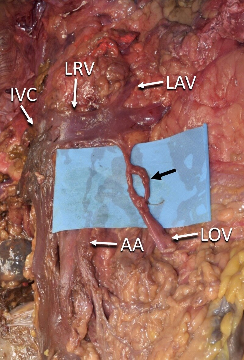
A fenestration in the left ovarian vein was found in a fresh-frozen female cadaver. The opening did not have any vessels or additional anatomical structures passing through it. The ovarian vein is also referred to as the female gonadal vein. This type of anatomical variation is clinically relevant in procedures that deal with the manipulation of the gonadal veins, specifically conditions such as ovarian vein thrombosis, ovarian vein stenosis, and pelvic congestion syndrome.

KEYWORDS:

anatomy; dissection; gonadal vein; obstetrics and gynecology; ovarian vein

The Current Trend of the Translational Research Paradigm.

Cureus. 2018 Mar 19;10(3):e2340. doi: 10.7759/cureus.2340.

Choi PJTubbs RSOskouian RJ.


Abstract

The translational research paradigm is a process of discovering basic science concepts and applying the knowledge in clinical practice, aiming to improve patient care. The stages involved in the paradigm form a complex network of shared knowledge amongst research collaborators, including patients. This nature of the paradigm allows those involved to work together effectively. However, the translational research paradigm is often overlooked by many scientists, educators, and research institutions. Hence, a large amount of comprehensive and hugely invested research projects fail to make a scientific impact. We aim to outline and describe this paradigm in order to aid in the successful translation of effective research.

KEYWORDS:

bi-directionality; community outreach; interdisciplinary training; multi-disciplinary; patient care; translational research

Emerging Cellular Therapies for Glioblastoma Multiforme.

Cureus. 2018 Mar 11;10(3):e2305. doi: 10.7759/cureus.2305.

Choi PJTubbs RSOskouian RJ.


Abstract

Glioblastoma multiforme (GBM) is the most common type of malignant primary brain cancer in adults. It is composed of highly malignant cells that display metastatic and angiogenic characteristics, making it resistant to current first-line chemotherapy with temozolomide, an alkylating agent. Despite many years of research, GBM remains poorly responsive to multiple available therapies, giving GBM patients, who receive the conventional combination of chemoradiotherapies and surgical resection, a dismal prognosis. There is growing evidence that the conventional systemic chemotherapeutic agents for GBM are ineffective in improving the disease progression. We aim to explore the emerging cellular therapies which may play a significant role in treating GBM.

KEYWORDS:

cellular therapy; cellular vehicle; glioblastoma multiforme; molecular envelope technology; oncolytic virus; temozolomide

A new superficial landmark for the odontoid process: a cadaveric study.

Anat Sci Int. 2018 Mar;93(2):203-206. doi: 10.1007/s12565-017-0390-1. Epub 2017 Feb 7.

Fisahn CMontalbano MJIwanaga JMoisi MLoukas MChapman JROskouian RJTubbs RS.


Abstract

When image guidance is not available or when there is a need to confirm the findings of such technology, superficial landmarks can still play a role in providing surgeons with estimations of the position of deeper anatomical structures. To our knowledge, surface landmarks for the position of the odontoid process have not been investigated. We have therefore performed an anatomical study to investigate such a landmark. One-centimeter metallic rods were placed on the philtrum of the upper lip of 20 cadaveric head specimens. To assess the position relative to the odontoid process, we took lateral and anteroposterior radiographs and recorded the measurements. Descriptive findings from radiographic observations indicated a reasonable approximation between the philtrum and the midpoint of the odontoid process. Based on our results, we suggest that the philtrum of the upper lip can serve as a first line estimation of the position of the odontoid process and can assist in verifying this bony structure following the use of image guidance.

KEYWORDS:

Anatomy; Axis; Cervical spine; Landmarks; Odontoid process; Philtrum

Highlights from the First Annual Spinal Navigation, Emerging Technologies and Systems Integration Meeting.

Abstract

This paper provides a detailed report of the "First Annual Spinal Navigation, Emerging Technologies and Systems Integration" meeting held December 3, 2016 at the Seattle Science Foundation.

KEYWORDS:

Spinal navigation; emerging technologies; robotics; spine; system integration; trauma; workflow

Rheumatoid arthritis in review: Clinical, anatomical, cellular and molecular points of view.

Clin Anat. 2018 Mar;31(2):216-223. doi: 10.1002/ca.22980. Epub 2017 Oct 27.

Sharif KSharif AJumah FOskouian RTubbs RS.


Abstract

Rheumatoid arthritis (RA) is the most common chronic autoimmune disease of the joints affecting close to 0.5-1.0% of the general population. Although the etiopathogenesis of RA remains elusive, the involvement of dendritic cells and type 17 T-helper cells appears to be pivotal in maintaining a state of chronic inflammation. RA is generally characterized by small joint involvement. A chronic inflammatory process leads to joint destruction and to tendon and ligament laxity and disintegration. These processes result in an imbalance of forces acting on the joints causing joint deformities including swan neck deformity, boutonniere deformity of the hands, flexion deformity of the wrist, lesser toe deformities, and others. In some instances, bony erosions subsequent to the RA disease process can result in life-threatening events including, for example, atlanto-axial subluxation, which can cause myelopathy and paralysis; and basilar invagination, which can cause brain stem injury and imminent death. Although less commonly involved, larger joints are not spared, as evidenced by the involvement of the elbow, hip, and shoulder joints in a sizable proportion of RA patients. The progression and prognosis of this disease entity are variable, guarded and dependent on the efficacy and response to treatment modalities employed. Inadequate management results in disease progression, which ultimately leads to joint erosion, destruction, deformities and substantial decrease in the functional quality of life. Clin. Anat. 31:216-223, 2018.

© 2017 Wiley Periodicals, Inc.

KEYWORDS:

Rheumatoid arthritis; anatomy; arthritis; arthrology; autoimmune disease; complications; immunopathology; inflammatory; joints; medicine; pannus

Anatomical variations; Classification; Clinical significance; Extensor digitorum brevis manus; Extensor indicis proprius; Forearm

Clin Anat. 2018 Mar;31(2):202-215. doi: 10.1002/ca.22939. Epub 2017 Dec 4.

Shoja MMJohal JOakes WJTubbs RS


Abstract

Although the Chiari malformations are well-studied and described developmental anomalies, there remains some incongruity in regards to their underlying etiologies. A number of theories have been proposed with the purpose of accounting for the embryology and pathogenesis of the Chiari I and II malformations and their associated complications and clinical syndromes. The present review aims to review the pertinent literature for all of the main theories that have been proposed, and outline their validity and relevance to our contemporary understanding of these anomalies. Clin. Anat. 31:202-215, 2018.

© 2017 Wiley Periodicals, Inc.

KEYWORDS:

Arnold-Chiari malformation; cerebrospinal fluid pressure; etiology; hydrocephalus; mechanism; syringomyelia

Extensor indicis proprius muscle and its variants together with the extensor digitorum brevis manus muscle: a common classification. Clinical significance in hand and reconstructive surgery.

Surg Radiol Anat. 2018 Mar;40(3):271-280. doi: 10.1007/s00276-018-1981-9. Epub 2018 Jan 30.

Georgiev GPTubbs RSIliev AKotov GLandzhov B.


Abstract

PURPOSE:

The extensor indicis proprius (EIP) is a muscle of the forearm that originates from the posterior surface of the ulna and the adjacent interosseous membrane and attaches to the index finger. Many anatomical variations of this muscle have been reported in the literature. The extensor digitorum brevis manus (EDBM) is a variant muscle found on the back of the wrist and hand. These muscle variants should be considered in the context of clinical syndromes and reconstructive hand surgery. The aim of the present study was to describe in detail the normal and variant anatomy of these muscles and propose new systematic classifications.

MATERIALS AND METHODS:

Formol-carbol-fixed Europid upper limbs (n = 176) were examined in the Department of Anatomy, Histology and Embryology at the Medical University of Sofia for variations of the EIP and occurrences of the EDBM.

RESULTS:

We found normal anatomy in 147 (84%) of the specimens examined and we measured the length and width of the muscle belly and tendon of the EIP. Variations of the EIP in the other 29 upper limbs (16%) included replacement of the EIP by an EIB, coexistence of the EIP and EIB, presence of accessory tendons, and additional muscles.

CONCLUSIONS:

Knowledge of anatomical variations of forearm muscles is important because they can cause clinical syndromes or have implications for reconstructive surgery, most often as tendon transfers in response to functional loss of other forearm muscles.

KEYWORDS:

Anatomical variations; Classification; Clinical significance; Extensor digitorum brevis manus; Extensor indicis proprius; Forearm

Prevalence of Petrosquamosal Sinus and Its Clinical Significance: Radiologic Study and Meta-analysis.

World Neurosurg. 2018 Mar;111:e616-e623. doi: 10.1016/j.wneu.2017.12.132. Epub 2017 Dec 30.

Pękala PARybus JJStachura MPękala JRSkinningsrud BTubbs RSTomaszewski KAHenry BM.


Abstract

OBJECTIVE:

The petrosquamosal sinus (PSS) is clinically important as it represents a potential hemorrhagic hazard during neurosurgical procedures. This double-approach radiologic study and meta-analysis aimed to assess the imaging characteristics of the PSS on computed tomography (CT) and systematically analyze its prevalence to improve imaging accuracy and assist in neurosurgical management.

METHODS:

The retrospective radiologic study included CT scans of 388 patients aged 18 years or older. All patients with any visible intracranial pathologies or history of head trauma were excluded. To identify articles eligible for inclusion in our meta-analysis, an extensive search was conducted through PubMed, Embase, ScienceDirect, Web of Science, and CNKI. Data extracted included investigative method, prevalence of the PSS among subjects, ethnicity, gender, side, laterality, and mean diameter. The extracted data were pooled into a meta-analysis with a random-effects model.

RESULTS:

The radiologic study showed that the PSS was visualized in 25.5% of the sides examined, that it was slightly more common among females (27.5%) than males (23.8%) and on the left (21.7%) than the right (29.4%). Its mean diameter was 1.14 ± 0.58 mm. The meta-analysis revealed that the overall prevalence of the PSS in the population is 11.1%, that it is more common in females (27.4%) than males (17.6%), and that a unilateral PSS is more common (78.2%) than a bilateral one (21.8%).

CONCLUSIONS:

As a PSS is present in about one tenth of the population, it is important to consider the possibility that it is present when undertaking neurosurgical procedures and to assess preoperative CT scans to avoid complications.

Copyright © 2017 Elsevier Inc. All rights reserved.

KEYWORDS:

Anatomy; Evidence-based anatomy; Meta-analysis