An Unusual Variation of the Accessory Nerve.

Cureus. 2018 Jun 9;10(6):e2774. doi: 10.7759/cureus.2774.

Alimi YIwanaga JLoukas MOskouian RJTubbs RS.


Abstract

The accessory nerve is an important nerve in the head and neck regions. Some variants of this nerve's anatomy have been reported. Herein, we present an unusual report and review the extant medical literature regarding other more commonly found derailments of this nerve's anatomy.

KEYWORDS:

accessory nerve; internal jugular vein; sternocleidomastoid muscle; variations

A Comprehensive Review of the Fabella Bone.

Cureus. 2018 Jun 5;10(6):e2736. doi: 10.7759/cureus.2736.

Dalip DIwanaga JOskouian RJTubbs RS.


Abstract

The fabella is a sesamoid bone that is embedded in the lateral head of the gastrocnemius muscle and often articulates directly with the lateral femoral condyle. It is present in 10-30% of the general population with a higher incidence in Asians. The fabella can lead to various pathologies such as fabella pain syndrome and common fibular nerve palsy. Conservative treatment involves physical therapy or injecting local anesthetics or steroids around this bone. However, if symptoms persist, then a fabellectomy can be performed. Physicians should be aware of the fabella bone and the multiple pathologies associated with it in order to provide the best treatment and management for patients.

KEYWORDS:

anatomy; fabellectomy; femoral condyle; gastrocnemius; knee pain; sesamoid; shock wave therapy; variations

The influence of ancient Greek thought on fifteenth century anatomy: Galenic influence and Leonardo da Vinci.

Childs Nerv Syst. 2018 Jun;34(6):1095-1101. doi: 10.1007/s00381-017-3462-6. Epub 2017 May 29.

Tubbs RIGonzales JIwanaga JLoukas MOskouian RJTubbs RS.


Abstract

Leonardo da Vinci (1452-1519) can be called one of the earliest contributors to the history of anatomy and, by extension, the study of medicine. He may have even overshadowed Andreas Vesalius (1514-1564), the so-called founder of human anatomy, if his works had been published within his lifetime. While some of the best illustrations of their time, with our modern knowledge of anatomy, it is clear that many of da Vinci's depictions of human anatomy are inaccurate. However, he also made significant discoveries in anatomy and remarkable predictions of facts he could not yet discover with the technology available to him. Additionally, da Vinci was largely influenced by Greek anatomists, as indicated from his ideas about anatomical structure. In this historical review, we describe da Vinci's history, influences, and discoveries in anatomical research and his depictions and errors with regards to the musculoskeletal system, cardiovascular system, nervous system, and other organs.

Guest UserComment
Anatomical Examination of Mandibular Condyle Protrusion Into the Middle Cranial Fossa.

J Craniofac Surg. 2018 Jun;29(4):1069-1071. doi: 10.1097/SCS.0000000000004425.

Wilson CIwanaga JOskouian RJTubbs RS.


Abstract

Trauma to the mandible can occasionally be a life-threatening event. Although extremely rare with only 56 reported patients in the English language, fracture of the floor of the middle cranial fossa with protrusion of the mandibular condyle into the middle cranial fossa was first reported in 1963 by Dingman. The authors review the anatomy of the temporomandibular joint in relation to the middle cranial fossa and demonstrate the possible complications due to condylar intrusion with anatomical dissection.

Guest UserComment
The retromolar canals and foramina: radiographic observation and application to oral surgery.

Surg Radiol Anat. 2018 Jun;40(6):647-652. doi: 10.1007/s00276-018-2005-5. Epub 2018 Mar 29.

Kikuta SIwanaga JNakamura KHino KNakamura MKusukawa J.


Abstract

PURPOSE:

The retromolar canal (RMC) and foramen (RMF) are anatomic variants in the retromolar area of the mandible. The purpose of this study was to clarify the relationship between the RMC and RMF and related complications, and to reveal how the RMC could impact the mandibular anatomy using cone-beam computed tomography (CBCT) and panoramic images (PAN).

MATERIALS AND METHODS:

CBCT and PAN images of 50 patients were retrospectively analyzed to investigate the morphology of the RMC and RMF, and their impact on impacted third molar surgery and orthognathic surgery.

RESULTS:

In PAN images, neither the RMC nor RMF was detected. In CBCT images, the RMCs were detected in 26% (13/50) of the patients. A double RMC was detected on one side of one patient. The diameter of the RMC ranged from 0.8 to 2.9 mm (mean; 1.5 ± 0.6 mm), and the RMF ranged from 0.6 to 2.3 mm (mean; 1.1 ± 0.5 mm). No patients experienced unexpected bleeding. Unilateral postoperative hypoesthesia of the buccal gingiva in the molar region was reported in 6.7% of patients with the RMC.

CONCLUSIONS:

Hypoesthesia of the buccal gingiva in the lower molar region may be the main complication when the RMC is damaged.

KEYWORDS:

Anatomic variation; Cone-beam computed tomography; Mandible; Oral surgery; Retromolar canal; Retromolar foramen

Coracobrachialis Longus Muscle: Humeroepitrochlearis.

Cureus. 2018 May 13;10(5):e2615. doi: 10.7759/cureus.2615.

Georgiev GPTubbs RSLandzhov B.


Abstract

During a routine anatomical dissection of the right brachium of a 68-year-old male cadaver, an extremely rare variation of the coracobrachialis longus muscle was discovered. It started from the medial surface of the middle part of the humerus with a well-formed muscle portion and then continued into the well-presented distal tendinous portion, which was attached to the medial epicondyle of the humerus. We also briefly review the reported variations of the coracobrachialis and their potential clinical significance.

KEYWORDS:

coracobrachial variations; humeroepitrochlearis muscle

Variant Distal Ulnar Nerve Loop: A Previously Undescribed Anatomical Finding.

Cureus. 2018 May 10;10(5):e2604. doi: 10.7759/cureus.2604.

Patel MIwanaga JOskouian RJTubbs RS.


Abstract

A previously undescribed variant ulnar nerve loop was discovered during the routine anatomic forearm and hand dissection of an adult female. The major finding was that of a large loop traveling around the distal tendon of the flexor carpi ulnaris. The variation presented here appears to be unique. The exact function of such derailed anatomy is not clear but, if found during surgery, might confound normal dissection methods or, when present, could result in varied clinical presentations regarding the sensory or motor examination of the hand.

KEYWORDS:

anatomy; anomalies; cadaveric; hand surgery; loop; ulnar nerve; variations

Consensus guidelines for the uniform reporting of study ethics in anatomical research within the framework of the anatomical quality assurance (AQUA) checklist.

Clin Anat. 2018 May;31(4):521-524. doi: 10.1002/ca.23069. Epub 2018 Mar 25.

Henry BMVikse JPekala PLoukas MTubbs RSWalocha JAJones DGTomaszewski KA.


Abstract

Unambiguous reporting of a study's compliance with ethical guidelines in anatomical research is imperative. As such, clear, universal, and uniform reporting guidelines for study ethics are essential. In 2016, the International Evidence-Based Anatomy Working group in collaboration with international partners established reporting guidelines for anatomical studies, the Anatomical Quality Assurance (AQUA) Checklist. In this elaboration of the AQUA Checklist, consensus guidelines for reporting study ethics in anatomical studies are provided with in the framework of the AQUA Checklist. The new guidelines are aimed to be applicable to research across the spectrum of the anatomical sciences, including studies on both living and deceased donors. The authors hope the established guidelines will improve ethical compliance and reporting in anatomical research. Clin. Anat. 31:521-524, 2018.

© 2018 Wiley Periodicals, Inc.

KEYWORDS:

anatomy; ethics; methodology; reporting guidelines; research

Embryology of the craniocervical junction and posterior cranial fossa, part I: Development of the upper vertebrae and skull.

Clin Anat. 2018 May;31(4):466-487. doi: 10.1002/ca.23049. Epub 2018 Mar 9.

Shoja MMRamdhan RJensen CJChern JJOakes WJTubbs RS.


Abstract

Although the embryology of the posterior cranial fossa can have life altering effects on a patient, a comprehensive review on this topic is difficult to find in the peer-reviewed medical literature. Therefore, this review article, using standard search engines, seemed timely. The embryology of the posterior cranial fossa is complex and relies on a unique timing of various neurovascular and bony elements. Derailment of these developmental processes can lead to a wide range of malformations such as the Chiari malformations. Therefore, a good working knowledge of this embryology as outlined in this review of its bony architecture is important for those treating patients with involvement of this region of the cranium. Clin. Anat. 31:466-487, 2018.

© 2018 Wiley Periodicals, Inc.

KEYWORDS:

Chiari malformation; anatomy; hindbrain; spine

Anatomical study of the so-called "retromolar gland": Distinguishing normal anatomy from oral cavity pathology.

Clin Anat. 2018 May;31(4):462-465. doi: 10.1002/ca.23047. Epub 2018 Feb 22.

Iwanaga JNakamura KAlonso FKirkpatrick COskouian RJWatanabe KTubbs RS.


Abstract

The minor salivary glands in the retromolar trigone have rarely been studied. The aim of this study was to better define the anatomy of the minor salivary glands in the retromolar trigone and establish the relationships between these and adjacent structures. The gland in the retromolar trigone was exposed and its relationships to surrounding structures were observed on 20 cadaveric sides. The boundaries of the gland included the superior pharyngeal constrictor muscle, the tendon of the buccinator muscle, and loose connective tissue. The gland was not continuous with the pterygomandibular or parapharyngeal spaces, but loose connective tissue was present between glands in the retromolar trigone and the medial pterygoid muscle. To our knowledge, this is the first study to describe the detailed anatomy of the minor salivary gland in the retromolar trigone. We suggest that the minor salivary gland in the retromolar trigone should be named the "retromolar gland." Clin. Anat. 31:462-465, 2018.

© 2018 Wiley Periodicals, Inc.

KEYWORDS:

anatomy; mandible; metastasis; minor salivary glands; tumor

Embryology of the craniocervical junction and posterior cranial fossa, part II: Embryogenesis of the hindbrain.

Clin Anat. 2018 May;31(4):488-500. doi: 10.1002/ca.23048. Epub 2018 Mar 9.

Shoja MMRamdhan RJensen CJChern JJOakes WJTubbs RS.


Abstract

Although pathology of the hindbrain and its derivatives can have life altering effects on a patient, a comprehensive review on its embryology is difficult to find in the peer-reviewed medical literature. Therefore, this review article, using standard search engines, seemed timely. The embryology of the hindbrain is complex and relies on a unique timing of various neurovascular and bony elements. Derailment of these developmental processes can lead to a wide range of malformations such as the Chiari malformations. Therefore, a good working knowledge of this embryology as outlined in this review of the hindbrain is important for those treating patients with involvement of this region of the central nervous system. Clin. Anat. 31:488-500, 2018.

© 2018 Wiley Periodicals, Inc.

KEYWORDS:

Chiari; anatomy; brain stem; brain, nervous system; cerebellum; genetics; malformation

Distribution of the internal nasal branch of the infraorbital nerve to the nasal septum: Application to rhinoplasty.

J Plast Reconstr Aesthet Surg. 2018 May;71(5):665-669. doi: 10.1016/j.bjps.2017.12.004. Epub 2017 Dec 13.

Iwanaga JWatanabe KOskouian RJTubbs RS.


Abstract

BACKGROUND:

The course of the nerves along the nasal septum has not been clearly studied, and surgical procedures such as rhinoplasty require a more detailed topography of the nerve supply inside the septum. Therefore, we aimed to investigate the distribution of the internal nasal branch of the infraorbital nerve inside the nasal septum and to define the relationship between it and the nasal cartilages.

METHODS:

Fourteen sides from eight fresh frozen and embalmed Caucasian cadaveric heads were dissected. The specimens were derived from three males and five females. The ages of the cadavers at death ranged from 65 to 84 years. The course of the internal nasal branch and its relationship between the nasal cartilages were observed using a surgical microscope.

RESULTS:

On all sides, the internal nasal branch approached the medial crus of the major alar cartilage from behind and traveled anteriorly below the medial crus of the major alar cartilage while giving off anterior inferior septal, middle inferior septal, and posterior inferior septal branches.

CONCLUSIONS:

Based on the results of this study, we suggest that procedures of the nasal cavity such as rhinoplasty could be modified to avoid injuring the main trunk of the internal nasal branch of the infraorbital nerve inside the nasal septum.

Copyright © 2017 British Association of Plastic, Reconstructive and Aesthetic Surgeons. Published by Elsevier Ltd. All rights reserved.

KEYWORDS:

Anatomy; Cosmetic surgery; Nasal septum; Nose; Rhinoplasty; Trigeminal nerve

Anatomic Study of Nutrient Foramina of Posterior Axis with Application to C2 Pedicle Screw Placement.

World Neurosurg. 2018 May;113:e408-e413. doi: 10.1016/j.wneu.2018.02.043. Epub 2018 Feb 14.

Tawfik TIwanaga JYilmaz EChoi PWilson CSimonds EMarks JJHarkey HLOskouian RJTubbs RS.


Abstract

OBJECTIVE:

Pedicle screws placed into C2 necessitate a thorough understanding of this bone's unique anatomy. Although multiple landmarks and measurements have been used by surgeons, these are often varied in the literature with no consensus. Herein, we studied one recently proposed landmark using the nutrient foramina of the posterior aspect of C2 for pedicle screw placement.

METHODS:

On 19 (38 sides) C2 dry bone specimens, the presence, size, location, and distance from the midline of the nutrient foramina found at the junction between the isthmus and lamina were documented and measured. In addition, to discern the source of the artery entering such foramina, an injected adult cadaver was dissected.

RESULTS:

The number of foramina ranged from 0-5 with a mean of 1.84. On 3 sides, no foramina were identified. The mean diameter of the foramina was 0.57 mm. The location of the foramina was at position 1 on 9.5% of sides, position 2 on 66.4% of sides, and position 3 on 24.1% of sides. The mean horizontal distance from the midline of the spinous process of C2 to the foramina was 25.17 mm. In the cadaveric specimen, the source of the artery entering these C2 nutrient foramina was found to be distal branches of the deep cervical artery.

CONCLUSIONS:

We found the nutrient foramina of the C2 laminae are useful for pedicle screw placement. However, there are minor variations of the number and position of these structures. Lastly, on the basis of our study, 7.9% (n = 3) of sides will not have such foramina.

Copyright © 2018 Elsevier Inc. All rights reserved.

KEYWORDS:

Anatomy; Axis; Cervical spine; Landmarks; Pedicle screw

S2 Alar-Iliac Screw Insertion: Technical Note with Pictorial Guide.

World Neurosurg. 2018 May;113:e296-e301. doi: 10.1016/j.wneu.2018.02.009. Epub 2018 Feb 10.

Yilmaz E1, Abdul-Jabbar A2, Tawfik T2, Iwanaga J3, Schmidt CK3, Chapman J2, Blecher R2, Tubbs RS4, Oskouian RJ5.


Abstract

BACKGROUND:

The S2 alar-iliac (S2AI) screw is a modification of the iliac fixation technique using the space between the neuroforamina of S1 and S2 as an insertion point to fix the sacrum to the ilium. To our knowledge, an anatomic review of the S2AI technique has not been described and the insertion point is vague and angles differ in reports from the literature. The purpose of the current anatomic illustration is to provide step-by-step techniques with fluoroscopic imaging to help confirm the safe placement of S2AI screws.

METHODS:

The procedure was performed on the left and rights sides of a fresh, frozen, and thawed predissected male cadaver in a surgical training facility through a standard posterior midline exposure for placement of the S2AI screws. All screws were placed by a fellowship-trained spine surgeon and an attending spine surgeon.

RESULTS:

The specimen was placed prone, and a midline incision begun at the L4 or L5 spinous process. Using the anteroposterior and inlet views, the S1 dorsal sacral foramen, the S1 endplate, and the sacroiliac joint can be identified. The insertion point is 10 mm laterally between the S1 and S2 foramina and near to the sacroiliac joint. Aim toward the anterior inferior iliac spine is ensured by using a 30°-40° lateral angulation in the transverse plane and 20°-30° caudal angulation in the sagittal plane depending on the sacral angulation. Using lateral fluoroscopy, the acetabulum and greater sciatic notch can be identified and screw misplacement can be avoided. The screw length is measured and is usually between 60 and 90 mm (8- to 9-mm diameter). An elevator is used to identify the outer sacral cortex. Anteroposterior, obturator-outlet, and teardrop views are used to ensure correct screw insertion.

CONCLUSIONS:

Fluoroscopic guidance is crucial for optimal S2AI screw placement. Using the described technique allows a safe and correct insertion of the S2AI screw.

Copyright © 2018 Elsevier Inc. All rights reserved.

KEYWORDS:

Lumbopelvic fixation; S2AI; Sacrum; Spinopelvic fixation; Technique

The Leg Wound of King Philip II of Macedonia.

Cureus. 2018 Apr 18;10(4):e2501. doi: 10.7759/cureus.2501.

Brandmeir NPayne RRizk ETubbs RSArsuaga JLBartsiokas A.


Abstract

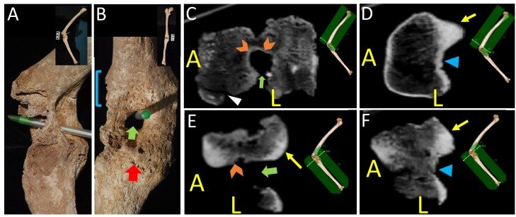
Objective King Philip II, the father of Alexander the Great, suffered a penetrating wound to the leg from a spear that left him severely handicapped. His skeletal remains represent the first and only case of an injury from ancient Greece that can be directly compared to its historical record. The objective of the study was to confirm the identity of the male occupant of Royal Tomb I in Vergina, Greece as Philip II of Macedonia by providing new evidence based on anatomical dissection and correlation with the historical description of the wounds. Methods Radiographs and photographs of the leg in Royal Tomb I in Vergina were examined. Anatomical dissection of a cadaver with a reconstructed wound similar to Philip's was also completed to identify associated soft-tissue injuries. Results The left leg was penetrated by an object at the knee which resulted in joint diastasis, external rotation of the tibia, knee ankylosis, and formation of a granuloma around the related object. This caused massive trauma to the joint but spared the popliteal artery. This resulted in ligamentous injury as well as injury to the peroneal nerve and probably the tibial nerve, resulting in a complete palsy of those nerves. Conclusion This evidence exactly matches the historical sources and shows conclusively that the leg and Tomb I belong to Philip II. The anatomic and archaeologic evidence also serve as independent verification of some of the historical record of that period, better enabling scholars to judge the reliability of various texts. Furthermore, it gives invaluable information about surgical practices in ancient Greece according to Hippocratic methods and their outcomes. Finally, this sheds new light on the occupants of Royal Tomb II including the fact that the armor recovered there may have belonged to Alexander the Great.

KEYWORDS:

alexander the great; joint granuloma; knee granuloma; peroneal nerve injury; philip ii

Cranial Nerve Foramina: Part II - A Review of the Anatomy and Pathology of Cranial Nerve Foramina of the Posterior Cranial Fossa.

Cureus. 2018 Apr 18;10(4):e2500. doi: 10.7759/cureus.2500..

Edwards BWang JMIwanaga JLoukas MTubbs RS.


Abstract

Cranial nerve foramina are integral exits from the confines of the skull. Despite their significance in cranial nerve pathologies, there has been no comprehensive anatomical review of these structures. Owing to the extensive nature of this topic we have divided our review into two parts; Part II, presented here, focuses on the foramina of the posterior cranial fossa and discusses each foramen's shape, orientation, size, surrounding structures, and structures that pass through it. Furthermore, by comparing foramen sizes against the cross-sectional areas of their contents, we determine the amount of free space available within each. We also review lesions that can obstruct each foramen and discuss the clinical consequences.

KEYWORDS:

cranial nerve; foramen; foramen magnum; hypoglossal canal; internal acoustic meatus; jugular foramen; posterior fossa; skull base

Treatment of Arrow Wounds: A Review.

Cureus. 2018 Apr 13;10(4):e2473. doi: 10.7759/cureus.2473.

Shereen ROskouian RJLoukas MTubbs RS.


Abstract

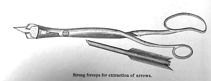
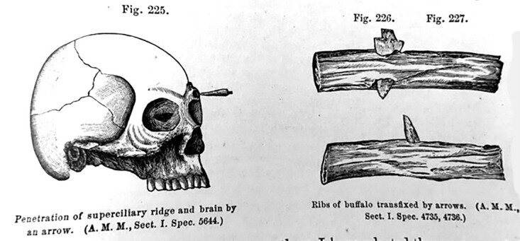
The arrow is one of the oldest weapons invented that has ties back to ancient civilization. With the advancement of modern weaponry, literature concerning the management of this traumatic wound has dwindled.  However, there are older written accounts that have led to our understanding of how we manage injuries inflicted by the arrow. One of the more comprehensive accounts was produced in the 19th century by a United States (US) Lieutenant Colonel named JH Bill. Recent cases in forensic pathology, as well as instances of trauma concerning arrowhead injuries, have documented a lack of comprehensive literature for the management of such injuries. Thus, the goal of our review is to evaluate the literature and provide a record of the different presentations, complications, and ways to manage arrow injuries.

KEYWORDS:

arrowhead; management; penetrating injury; trauma

Communication Between a Dorsal and Ventral Ramus: A Conflict for Traditional Anatomical Teaching.

Cureus. 2018 Apr 10;10(4):e2460. doi: 10.7759/cureus.2460.

Patel M1, Iwanaga J2, Oskouian RJ3, Tubbs RS4.


Abstract

Counterintuitive to anatomic organization is the communication between a dorsal ramus and a ventral ramus, which are traditionally taught as serving specific anatomical fields. Herein, we discuss the cadaveric findings of such an example of neural intercommunication between the dorsal and ventral rami and review the potential embryology. While these two nerves are complementary in their function, it is not understood as to the reason for such an anatomical connection that breaches normal anatomical "laws."

KEYWORDS:

anatomy; cadaver; cervical plexus; dissection; dorsal ramus; greater occipital nerve; lesser occipital nerve; spinal nerve; ventral ramus